|
An acquired, noninherited form of the disease may appear in patients with lymphoma, particularly Hodgkin's disease , but this form has been reported also in association with carcinoma and sarcoidosis. The expression of profilaggrin is reduced in this disorder. Profilaggrin mRNA in keratinocytes cultured from subjects with ichthyosis vulgaris is unstable and has a shorter half-life compared with that in normal cells. Therefore, a stabilizing factor may be absent or functionally inactive .
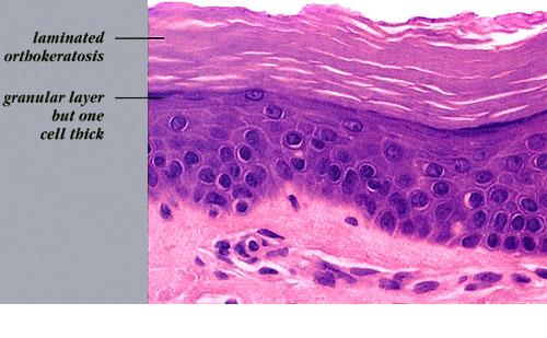
Histopathology.
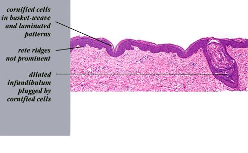
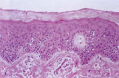
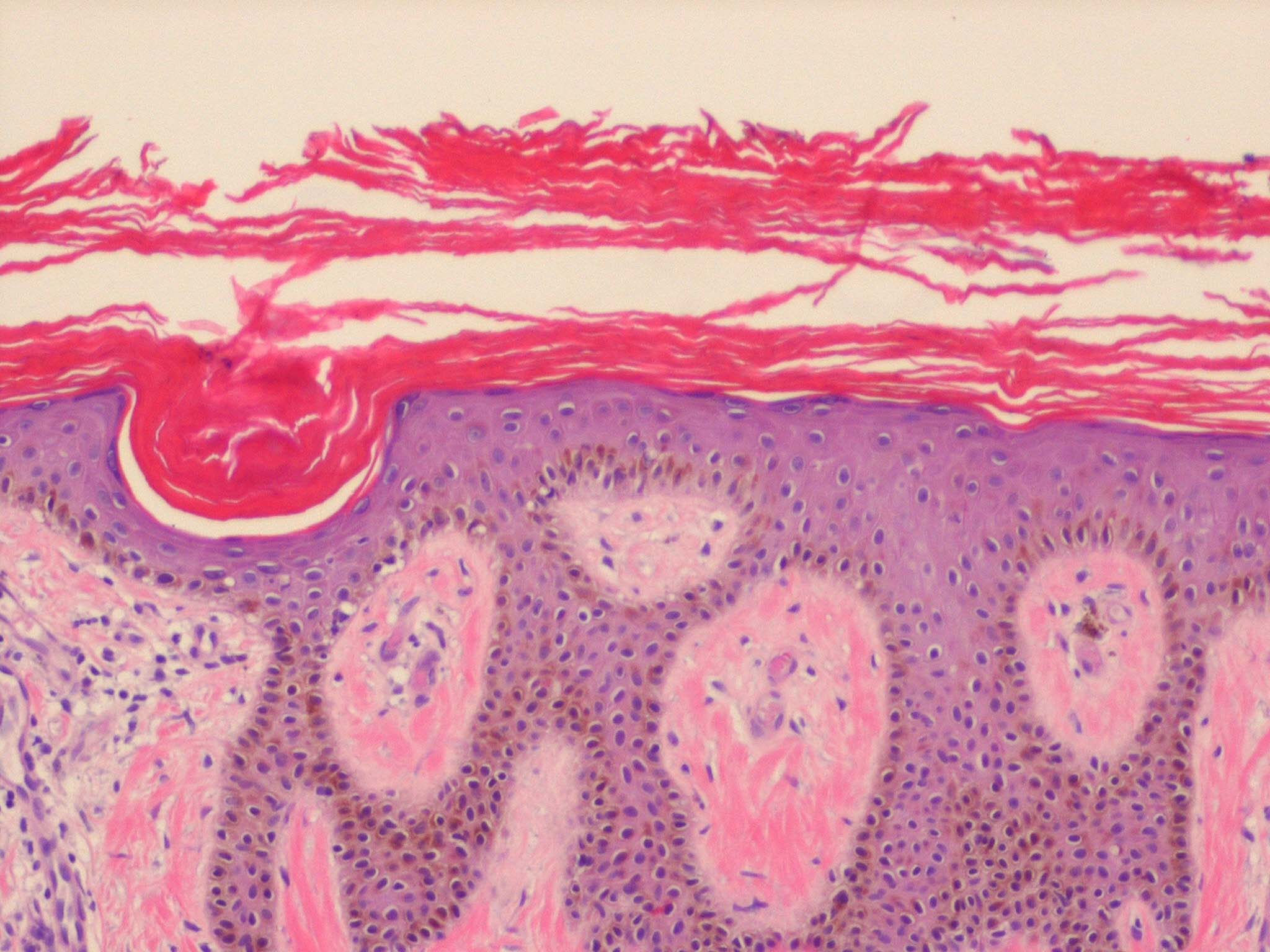
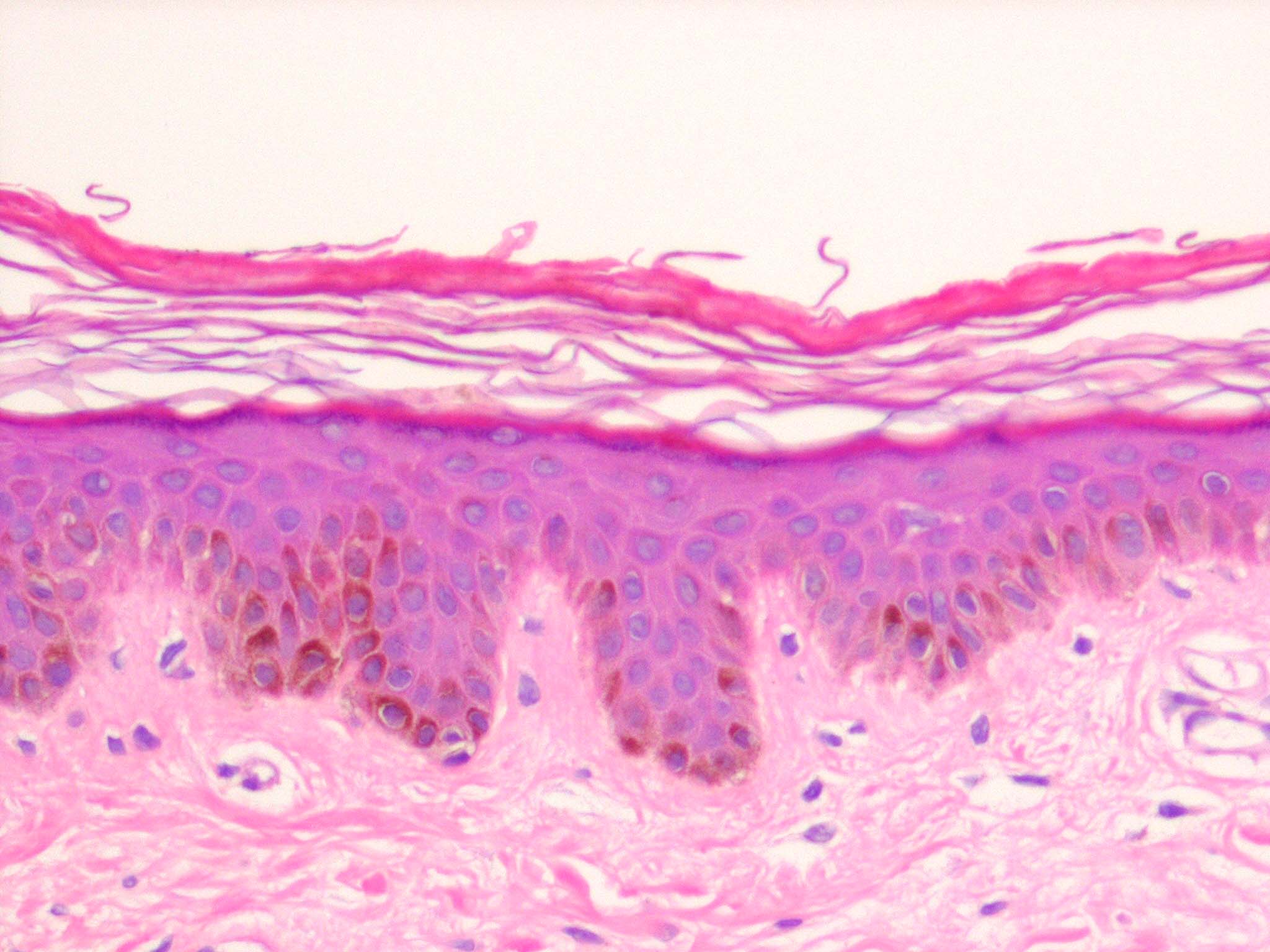
The characteristic finding is the association of a moderate degree of hyperkeratosis with a thin or absent granular layer . The hyperkeratosis often extends into the hair follicles, resulting in large keratotic follicular plugs. The dermis is normal.
Histogenesis.
Labeling with tritiated thymidine shows a normal rate of epidermal proliferation . The hyperkeratosis is regarded as a retention keratosis resulting from increased adhesiveness of the stratum corneum. The reason for this, as seen by electron microscopy, is a delay in the dissolution of the desmosomal disks in the homy layer. Keratohyaline granules are regularly seen on electron microscopy, in contrast to light microscopy. The stratum granulosum, however, consists of only a single layer, and the keratohyaline granules appear small and crumbly or spongy, which is evidence of defective synthesis. The reason for the inadequate formation of keratohyaline granules lies in a defect in the synthesis of filaggrin, a histidine-rich protein . Defective profilaggrin expression in ichthyosis vulgaris may be a result of selectively impaired posttranscriptional control . In noninherited ichthyosis vulgaris associated with neoplasia, the keratohyaline granules have been described as being small but showing a normal structure, indicating a reduced but not an abnormal synthesis
Differential Diagnosis.
Although the noninflamed but dry skin of patients with atopic dermatitis clinically resembles ichthyosis vulgaris, on histologic examination it does not show the features of ichthyosis vulgaris but rather increased epidermal thickness, patchy parakeratosis, and slight hypergranulosis in places, as seen in chronic dermatitis .
|