|
Hailey Hailey disease= الفقاع العائلي السليم هيلي هيلي |
 |
 |
 |
|
HaileyHailey Disease
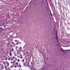
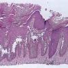
Familial benign pemphigus is inherited as an autosomal dominant trait, with a family history obtainable in about two thirds of patients. Genetic studies have localized the key mutations to the ATP2C1 gene on chromosome 3q , specifically 3q21-q24, the area responsible for ATP-dependent calcium transport . It is characterized by a localized, recurrent eruption of small vesicles on an erythematous base. By peripheral extension, the lesions may assume a circinate configuration. The sites of predilection are the intertriginous areas, especially the axillae and the groin. Only very few instances of mucosal lesions have been reported, of the mouth , the labia majora , and the esophagus .
|
|
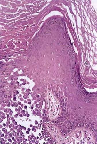
Histopathology.
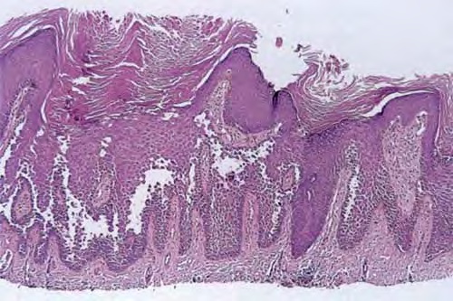
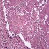
Although, as in Darier's disease, early lesions may show small suprabasal separations, so-called lacunae, fully developed lesions show large separations, that is, vesicles and even bullae, in a predominantly suprabasal position . Villi, which are elongated papillae lined by a single layer of basal cells, protrude upward into the bulla, and in some cases,
|
|
narrow strands of epidermal cells proliferate downward into the dermis. Many cells of the detached stratum malpighii show loss of their intercellular bridges; thus, acantholysis affects large portions of the epidermis.
|
|
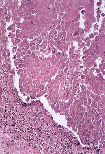
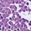
Individual cells and groups of cells usually are seen in large numbers in the bulla cavity. Despite the extensive loss of intercellular bridges, the cells of the detached epidermis in many places show only slight separation from one another, because a few intact intercellular bridges still hold them loosely together. This quite typical feature gives the detached epidermis the appearance of a dilapidated brick wall.
|
|
Differentiation of familial benign pemphigus from Darier's disease as a rule is not very difficult, because in Darier's disease, the suprabasal separations usually are smaller, thus appearing as lacunae rather than as bullae; acantholysis is less pronounced, being limited to the lower epidermis, especially the suprabasal region; and dyskeratosis consisting of the formation of corps ronds and grains is much more evident.
|
|
Pemphigus vulgaris often resembles familial benign pemphigus to a striking degree, and in some specimens, histologic differentiation of these two diseases may be impossible. As a rule, however, there is less extensive acantholysis in pemphigus vulgaris, limited largely to the suprabasal region, so the detached epidermis appears normal and lacks the appearance of a dilapidated brick wall, and more severe degeneration of the acantholytic cells within and near the bulla cavity. The presence of eosinophils in the bulla points toward a diagnosis of pemphigus vulgaris, but their absence does not rule it out. In case of doubt, immunofluorescence will decide the issue.
|
|
There used to be much discussion as to whether familial benign pemphigus represents a vesicular variant of Darier's disease. Two points in favor of the basic unity of the two diseases were stressed: the alleged simultaneous presence of both diseases in the same patient and the occurrence of corps ronds in both diseases. However, it has become apparent that patients described as having both diseases were either cases of Darier's disease with vesicular lesions or cases of familial benign pemphigus with the presence of corps ronds
. Evidence against a relationship is also shown by the fact that in affected families, always only one of the two diseases occurs and the genetic basis of the two diseases has been elucidated and are clearly different though related-the ATP2C1 gene on chromosome 3q is mutated in Hailey-Hailey , while Darier's disease is due to a mutation in the the ATP2A2 gene on chromosome 12 .
|
|
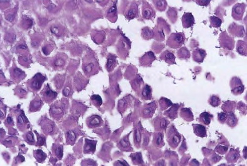
Many of the cells of the stratum malpighii that have lost all or most of their intercellular bridges show a fairly normal cytoplasm and a normal nucleus in which mitotic activity has even been observed . Some of the acantholytic cells, however, have a homogenized cytoplasm, suggesting premature partial keratinization. In some instances, such acantholytic cells with premature keratinization resemble the grains of Darier's disease. Occasionally, a few corps ronds are present in the granular layer .
|
|
Differential Diagnosis.
Histologically, familial benign pemphigus shares certain features with both Darier's disease and pemphigus vulgaris. In all three diseases, one finds predominantly suprabasal separation of the epidermis caused by acantholysis and resulting in lacunae or bullae and villi formation.
|
|
|
|
|