Digital Mucous Cyst
Digital mucous cysts (DMCs) are benign ganglion cysts of the digits, typically located at the distal interphalangeal (DIP) joints or in the proximal nail fold. They usually occur on the hands, although they have also been noted on the toes. The etiology of these cysts is uncertain but may involve mucoid degeneration. Often, these cysts are asymptomatic and do not require treatment. When treatment is indicated, medical therapies and surgical interventions of varying magnitudes may be attempted. Recurrence is common.
Historically, little attention has been directed at studying these cysts despite their frequency. In the literature, they have been referred to as cystomata, myxomatous cutaneous cysts, myxomatous degenerative cysts, periarticular fibromas, synovial lesions of the skin, periungual ganglions, mucous cysts, myxoid cysts, synovial cysts, dorsal cysts, nail cysts, cystic nodules, digital mucoid cysts, digital myxoid cysts, and digital mucinous pseudocysts.
Hippocrates first appreciated ganglion cysts, describing a knot of tissue full of fluid. In 1746, Eller concluded that ganglia formed from the herniation of the synovial lining of a joint. In 1882, Hyde first described the digital mucous cyst. In 1893, Ledderhose suggested that ganglia arose spontaneously in the subcutaneous tissue. In 1895, Ritschel proposed the earliest formulation of the theory that mucoid degeneration may be responsible for digital mucous cysts; Carp and Stout popularized the theory in 1928. Then, in 1947, Anderson reported that cysts caused the nail deformities.
The mechanism of formation of digital mucous cysts is unknown. Currently, it is believed that the cysts arise from mucoid degeneration of connective tissue and that this process, in most cases, involves communication with the adjacent DIP joint and possible coexistence of osteoarthritis. Clinical and radiographic evidence of osteoarthritis is common at the site of the cysts,1 and the frequent presence of osteophytes and spurring of the DIP joint were recognized in the 1970s. Active connection to the joint space may or may not exist, as the mucoblasts associated with the cyst appear capable of sustaining the process.
Ganglia are the most common tumor or cyst of the hand. They account for approximately 70% of all such tumors or cysts, with digital mucous cysts comprising 10-15% of the total.
.
Digital mucous cysts most often are asymptomatic and benign. Pain can result from the impingement of cysts on adjacent nerve fibers. Larger cysts can disfigure the affected digit. Nail deformities can occur.
Women are affected more often than men (female-to-male ratio of 2-2.5:1).
Digital mucous cysts usually occur in the fifth to seventh decades, yet they may be seen as early as the teenage years or among the elderly population. The mean age of onset is 60 years. One report describes a case in association with cutaneous mucinosis of infancy.
- Typically, the cysts are asymptomatic. They may appear suddenly or develop over a period of months. Grooving of the nail may precede the clinical manifestation of the cyst itself by up to 6 months. Often, osteoarthritis of the small joints is noted at the site of cyst emergence. Intermittent spontaneous discharge of cyst contents can occur, and, in a significant fraction of cases, cysts may disappear spontaneously.
- Antecedent trauma has been documented in a small minority of cases. As cysts enlarge, pain is an increasingly common complaint. Patients are also likely to complain about the appearance of larger cysts and may report interference with function.
Pertinent physical findings are limited to the skin, joints, and nail unit.
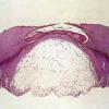
Digital mucous cyst at proximal nail fold.
- Skin - Primary lesion
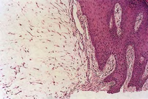
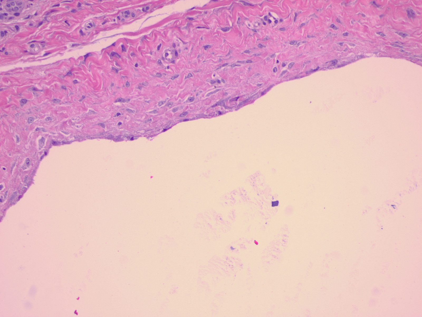
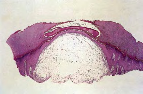
- Digital mucous cysts are usually solitary, round-to-oval, dome-shaped, firm-to-fluctuant papulonodules from 1-10 mm in diameter that have overlying skin that ranges from very thin to moderately thick.
- The cysts contain a viscous, gelatinous fluid that may be clear or yellow-tinged.
- Some cysts are verrucose.
- Pain is associated with relatively larger cysts.
- Skin - Distribution
- The cysts are located off the midline of the digits and, according to one series, are more common on the radial than ulnar aspect of the fingers.
- They most often are found on the dorsolateral aspect of the fingers, intradermally, between the DIP joint and proximal nail fold. Less frequently, they occur between the proximal nail fold and the nail plate, beneath the nail matrix, or in the pulp of the digit.
- Cysts most frequently are found on the middle or index finger of the dominant hand; toe involvement is less common.2
- Cysts located under the nail plate (subungual cysts) have common features that have been characterized in one series. In most cases, the lunula is discolored (most often red, less often blue) and transverse curvature of the nail is almost always increased, frequently resulting in lateral ingrowth.
- Skin - Color
- Digital mucous cysts are translucent to flesh-colored.
- When they are under the nail matrix, a red lunula and a longitudinal brownish band may be seen.
- Nails
- Longitudinal grooving or depression of the nail occurs when digital mucous cysts involve the posterior nail fold.
- Grooving may be accompanied by transverse ridging and thinning of the nail overlying the cyst.
- Gross disruption of the nail is less common.
- Digital mucous cysts are more likely to be above than below the nail matrix.
- Joints: A consensus has emerged that digital mucous cysts are frequently, if not always, located at osteoarthritic joints.
The causes of digital mucous cysts remain unclear. Historically, a variety of etiologies, including a tuberculous process, have been suggested. At present, it is believed that mucoid degeneration of connective tissue associated with proximal osteoarthritic changes is responsible for cyst formation. Trauma also may be a causative factor in some cases.