|
|
Androgenetic Alopecia
The hallmark of androgenetic alopecia (AGA) is follicular miniaturization, the process by which hair shafts become progressively finer and shorter. AGA results from an androgenic influence on hair follicles in certain areas of the scalp, and the pattern of involvement in the different sexes has led to use of the preferred terminology male-pattern or female-pattern hair loss. Some prefer this terminology over AGA because the etiologic role of androgens in women is not fully determined. AGA is the most common type of hair loss in males, affecting 50% of men by the age of 50 years and almost as many women, according to some reports , leading some to espouse that it is a physiologic process, not a pathologic one (283). AGA frequently shows a familial tendency, but inheritance does not follow simple Mendelian genetics and is most likely a polygenic trait
|
|
In male patients, the process usually begins with bifrontal thinning, often with similar changes over the vertex. In full expression of the condition, there may be almost total baldness of the entire frontal/parietal scalp. The most common pattern seen in women is one of diffuse thinning over the crown and frontal scalp with preservation of the frontal hairline. The process occurs with less severity in women, so that significant balding is unusual.
|
|
Histopathology.
Satisfactory evaluation of AGA can only be achieved from transverse sectioning of punch biopsy material , allowing for quantitative evaluation. Furthermore, samples smaller than 4 mm in diameter show an insufficient number of follicles for meaningful results.
|
|
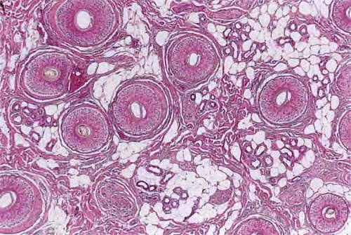
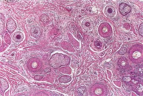
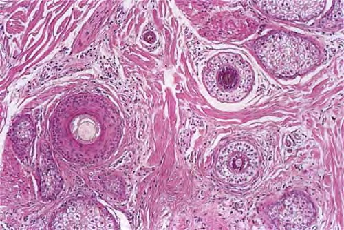
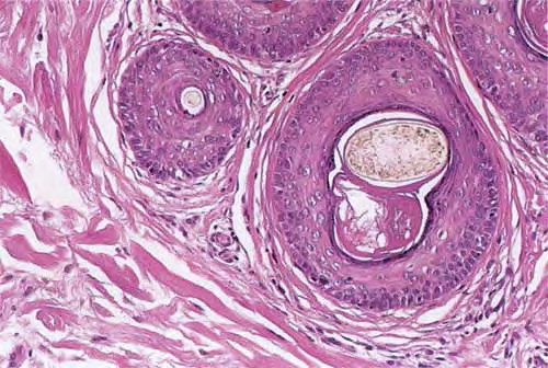
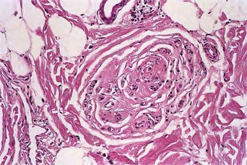
Diminution of follicular size, or miniaturization, is the histologic hallmark of AGA. To fully appreciate the number of miniaturized follicles, horizontal sections at the level of the lower infundibulum of terminal hair follicles should be examined because sections below this may miss the vellus hairs whose bulbs are situated in the upper dermis . Cursory inspection at low power reveals random variation in the caliber of hair follicles, consistent with a progressive miniaturization , and absence of well-defined follicular units . Although vellus hairs can be quickly identified by the fact that their shaft diameter is equal to or greater than the thickness of their inner root sheath, they are quantitatively defined as having a shaft diameter of 0.03 mm or less . In his 1984 paper that laid the groundwork for horizontal
|
|
interpretation of scalp biopsies, Headington also defined terminal hairs as having a shaft diameter of 0.06 mm or greater, thus recognizing an intermediate/indeterminate category in which the shaft diameter, i, measures 0.06 > i > 0.03 mm, between terminal and vellus hairs . An optical micrometer may be used to measure hair shaft diameter, if necessary.
|
|
However, slightly different definitions were used in subsequent articles on AGA, specifically with regard to the intermediate category. For example, Whiting classified terminal hairs as having a "shaft diameter exceeding 0.03 mm ... thicker than its inner root sheath," thereby grouping intermediate hairs with the terminal hair population . He then used a 3:1 (or less) terminal-to-vellus hair ratio as being diagnostic of male pattern AGA, although at other times he and others quoted a terminal-to-vellus hair ratio of less than or equal to 2: 1 to diagnose AGA . Whiting found the average terminal-to-vellus ratio in AGA to be 1.7:1 . Using similar definitions of terminal and vellus hairs, Sperling and Winton reported finding an average terminal-to-vellus ratio of 1 :6.1 , illustrating how, in advanced cases, miniaturized hairs outnumber terminal hairs (. In my opinion, intermediate
|
|
hairs should be grouped with the vellus hair population because more often they represent a transitional follicle undergoing miniaturization to a vellus hair (unless the patient is using agents such as minoxidil or finasteride to reverse the process); unfortunately, ratios for normal scalp and in AGA in which vellus and intermediate hairs are lumped together are not available in the literature and remain to be determined.
|
|
It is easy to undercount the number of vellus hairs, even with horizontal sections . Some vellus hairs may be so small as to be mistaken for keratinous debris. The use of toluidine blue stain was reported to be superior to the use of hematoxylin and eosin in identifying the tiniest of hairs because it stains the inner root sheath intensely blue .
|
|
The onset of AGA may be manifested by a telogen effluvium . Whiting found the telogen count in AGA to be 16.8%, as compared to 6.5% in controls (193), with telogen counts as high as 30% reported . Because the reduction in follicular size is associated with a shorter anagen phase (the smaller hairs do not grow as long), at a given point in time (for a histologic specimen this is the moment of biopsy harvest) a greater proportion of follicles will be found in telogen. There may be typical telogen hairs or, with increasing severity, telogen germinal units. A telogen germinal unit is the follicular epithelium that remains after the telogen hair shaft has been shed; it represents the secondary hair germ .Although vellus hairs continue to cycle through anagen and telogen in the papillary dermis, they are not considered to be part of the telogen count . Both miniaturization and increased conversion to telogen are associated with the finding of fewer terminal hair bulbs and more fibrous streamers in the deep dermis and subcutis . The fibrous streamers in AGA are thicker, more cellular, and possibly more fibrotic than those of normal follicles .
|
|
In advanced AGA there may be a reduced hair density with permanent loss of follicles, termed cicatricial pattern hair loss by Olsen and Olsen . Periinfundibular fibroplasia, ultimately leading to focal follicular scarring, may be responsible . In subtle cases, a second biopsy, taken from the occipital scalp, may be helpful for comparison because the occiput is typically uninvolved.
|
|
Lymphocytic infiltrates, defined as activated T cells in one study, are found around superficial vessels and in the vicinity of the lower infundibulum, sebaceous glands, and bulge in follicles transitioning to full miniaturization . The presence of inflammation has been reported in as many as 50% to 75% of cases and has been linked to peripilar signs seen clinically . However, mild perifollicular and upper dermal inflammation can be observed in normal scalp with a similar frequency to AGA and therefore is of no value in diagnosing AGA (. Moderate inflammation, however, was
|
|
seen more often in AGA than in normal individuals ; the infiltrate was composed primarily of lymphocytes and histiocytes, with rare neutrophils, plasma cells, and giant cells. It has been suggested that inflammation in AGA may result from Demodex, seborrheic dermatitis, actinic damage, cosmetics, and grooming agents and therefore should not be considered pathogenic . Nevertheless, its presence may have a bearing on the potential to respond to treatment; one study found that AGA patients with histologic inflammation or fibrosis showed less frequent regrowth on treatment with 2% minoxidil than AGA patients without inflammation , although the figures did not prove to be statistically significant. In conclusion, upper dermal inflammation can be seen in AGA, but it does not support or discount this diagnosis.
|
|
Pathogenesis
Androgens are the primary regulators of normal human hair growth. With the arrival of puberty, they
|
|
transform vellus hairs in many sites, such as the axilla, to terminal hairs, while at the same time mediating the opposite effect on certain scalp follicles in predisposed individuals . The effects of androgens on the hair follicle are mediated via androgen receptors in the hair papilla . The primary androgen responsible for these effects is dihydrotestosterone {DHT}, via androgen-receptor binding . Within the follicle, the enzyme 50reductase produces DHT from serum testosterone. Predisposed scalp displayed high levels of DHT and increased androgen-receptor expression in one study {29B}. In another study, frontal scalp follicles showed higher levels of androgen receptor and 50-reductase than occipital scalp follicles, and men exhibited higher levels than women .
|
|
A group of investigators found that the predominant form of 50-reductase in human scalp is type 1, concentrated in sebaceous glands . These same investigators found 50-reductase type 2 to be present in the inner layer of the outer root sheath, the proximal inner root sheath, and the infundibulum and some in sebaceous ducts. The type 2 isozyme is responsible for AGA. It is preferentially inhibited by finasteride, which decreases scalp production of DHT and promotes hair growth in males with AGA .
|
|
Other serum hormones, such as adrenal-derived dehydroepiandrosterone, may also be converted to DHT by some hair follicles, and steroid sulfatase found in the dermal papilla may be the responsible enzyme . This may explain why males affected by X-linked recessive ichthyosis due to a defect in steroid sulfatase do not develop AGA or do so only mildly {302}.
|
|
Apoptosis is an important part of hair follicle regression, and therefore factors that affect apoptosis may playa role in regulating the hair cycle . One study of apoptotic mechanisms in AGA suggested that levels of caspases, regulators of programmed cell death, and inhibitors of apoptosis may control hair follicle homeostasis . Another study found similar expression of bcl-2, p53, and other heat shock proteins in follicles from normal {occipital} scalp and follicles from scalp affected by AGA {frontal}, whereas follicular expression of the proliferation marker Ki-67 was decreased in AGA . The authors concluded that abnormal regulation of apoptosis was not involved in AGA, but a lower proliferation rate may be. This contradicts an analysis of bcl-2 and TUNEL {terminal deoxynucleotidyltransferase-mediated deoxyuridine triphosphate-biotin nick end-labeling} staining-markers of apoptotic activity-in scalp of cadaveric males with AGA, in which significant differences were noted between normal and alopecic scalp follicles ; the apoptotic "hot spot" was identified in the bulge-isthmus region.
Whiting postulated that the process of miniaturization cannot be explained simply by shortening of the anagen phase with repeated hair cycles. He suggested that miniaturization of some follicles occurred rapidly, over the course of a single hair cycle, possibly mediated by a change in the size of the dermal papilla, because vellus follicles have a smaller papilla than terminal follicles . In cell cultures of dermal papillae derived from balding scalp follicles, the cells were smaller and did not grow as well as those derived from nonbalding scalp follicles .
|
|
There is evidence that fibroplasia of the adventitial sheath surrounding the hair follicle is important to the pathogenesis of AGA . As previously noted, fibrous streamers in AGA may be more fibrotic than those of normal follicles . One study found Demodex to be present more frequently in patients with AGA and postulated that Demodex-induced inflammation could contribute to the process and possibly playa role in permanence {scarring} .
|
There exists a physiologic rest period during which the posttelogen follicle remains empty. This recently recognized phase of the hair cycle was termed kenogen . It may last longer and occur with greater frequency in AGA
|

