Incontinentia
Pigmenti.
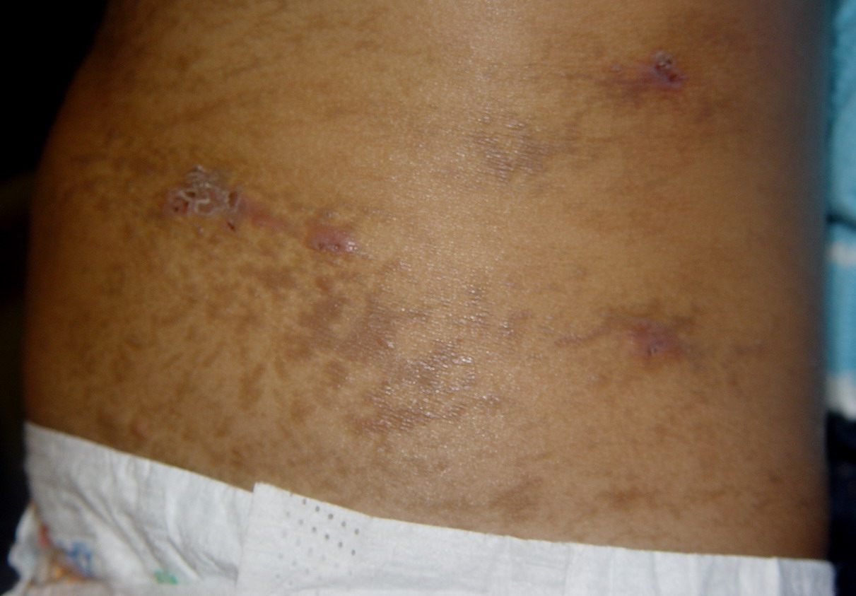
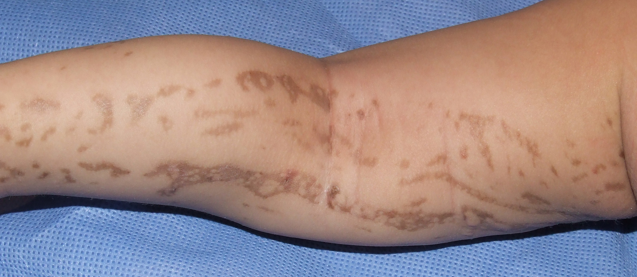
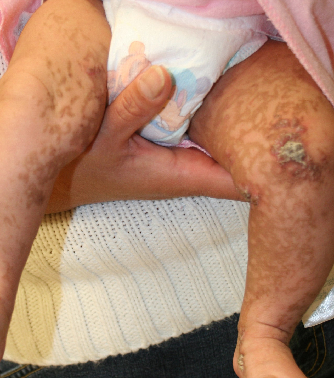
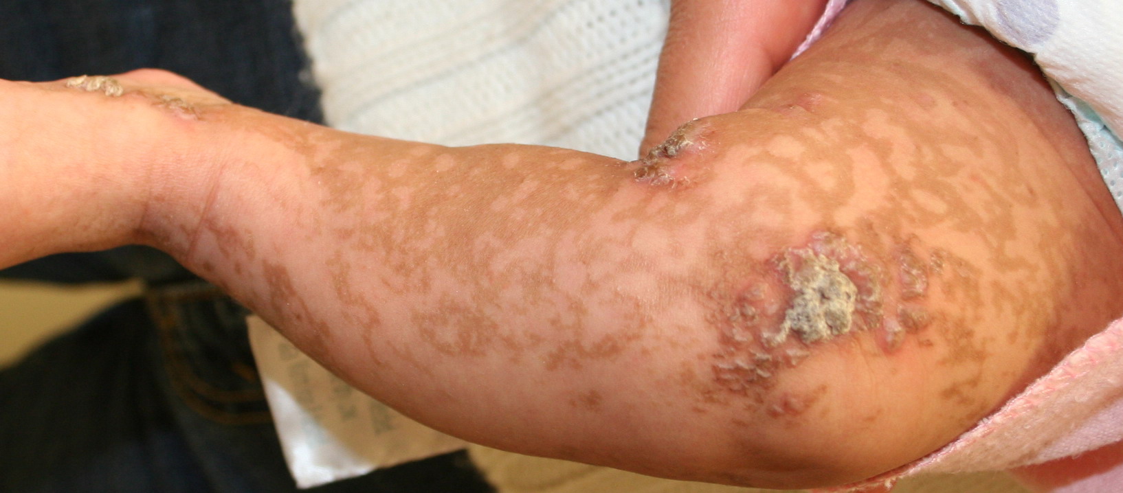
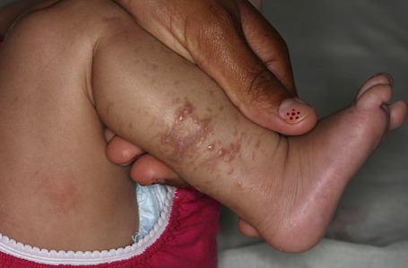
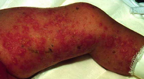
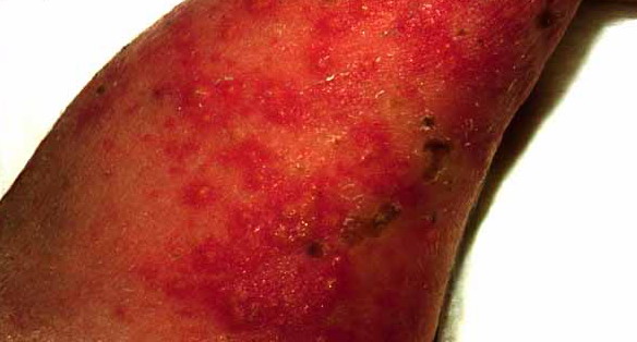
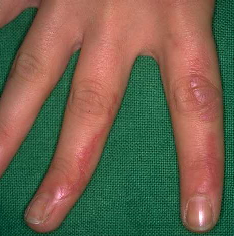
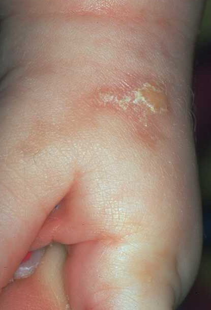
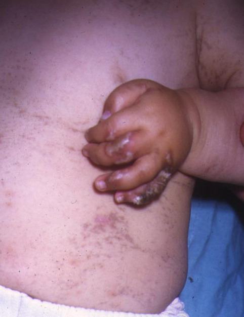
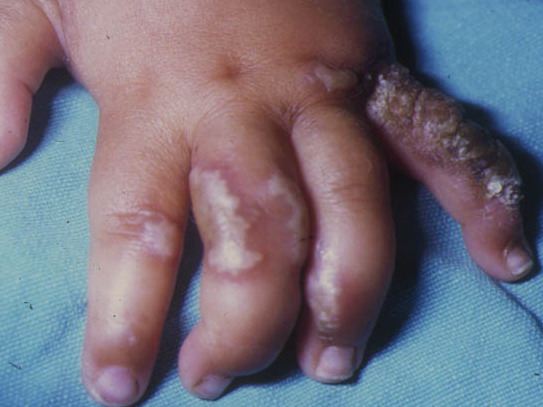
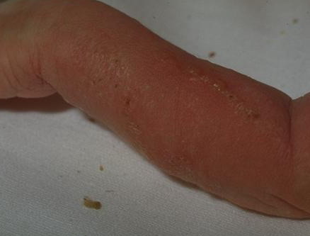
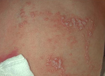
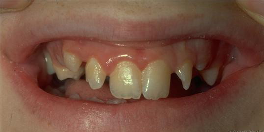
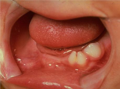
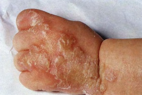
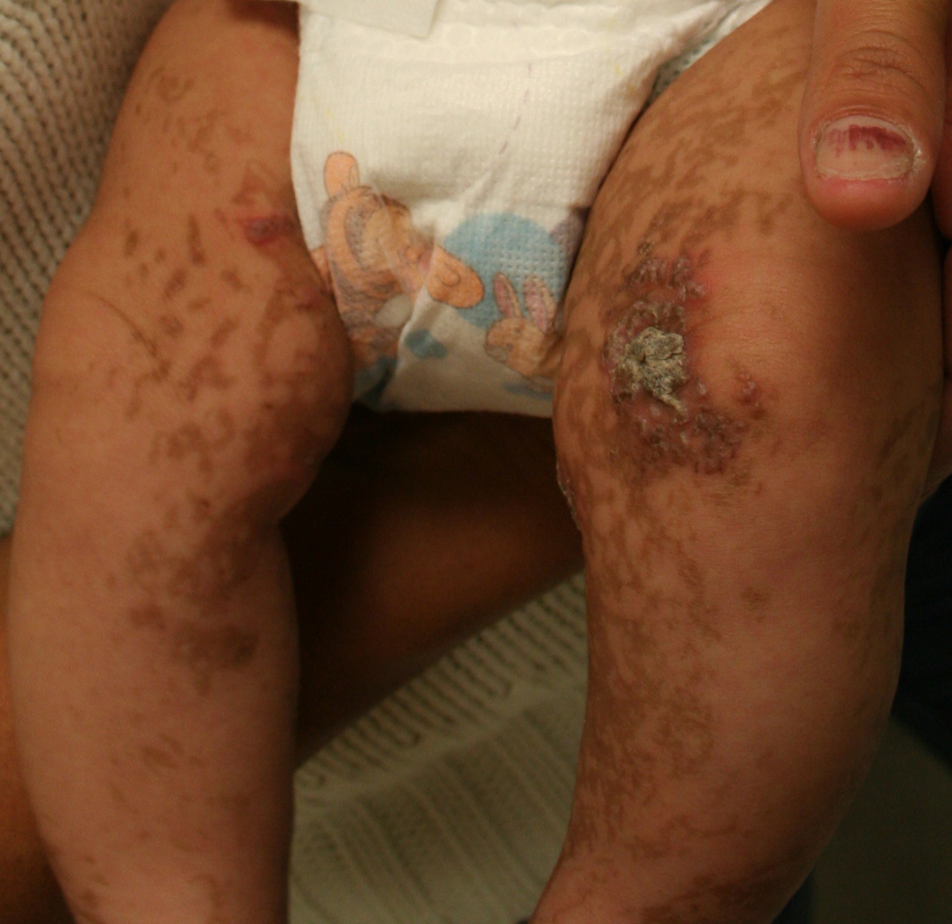
IP, also known as Bloch-Sulzberger syndrome, was first described by Garrod et al. in 1906. It is an X-linked, dominantly inherited disorder, reported primarily in females, and believed to be embryonic lethal in the majority of males. In most cases, IP is due to a mutation in a gene called NEMO [nuclear factor κB (NF-κB) essential modulator] in chromosome Xq28. It is characterized by typical skin lesions along the lines of Blaschko, which usually follow four cutaneous stages, sometimes with some overlap: (1) vesicular stage (from birth or shortly thereafter), (2) verrucous stage (between 2 and 8 weeks of age), (3) hyperpigmented stage (several months of age into adulthood), followed by (4) hypopigmentation stage (from infancy through adulthood) . A significant percentage of IP patients have ocular, dental, skeletal, and central nervous system anomalies.
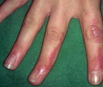
The cutaneous findings in the first stage represent the population of NEMO-deficient cells. The deficiency results in disruption of the signaling pathway, which results in failure to activate NF-κB, leading to apoptosis (NF-κB normally protects against tumor necrosis factor-induced apoptosis). The number of NEMO-deficient cells decreases secondary to apoptosis and is replaced by cells expressing the normal allele. Subsequently, the inflammatory and vesicular stage ends. The hyperproliferation in the second stage is likely due to the proliferation of normal NEMO keratinocytes. Hyperpigmentation subsequently appears in the third stage because of incontinence of melanin pigment from the epidermis into the dermis.
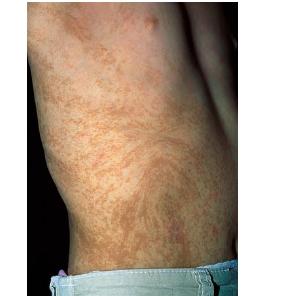
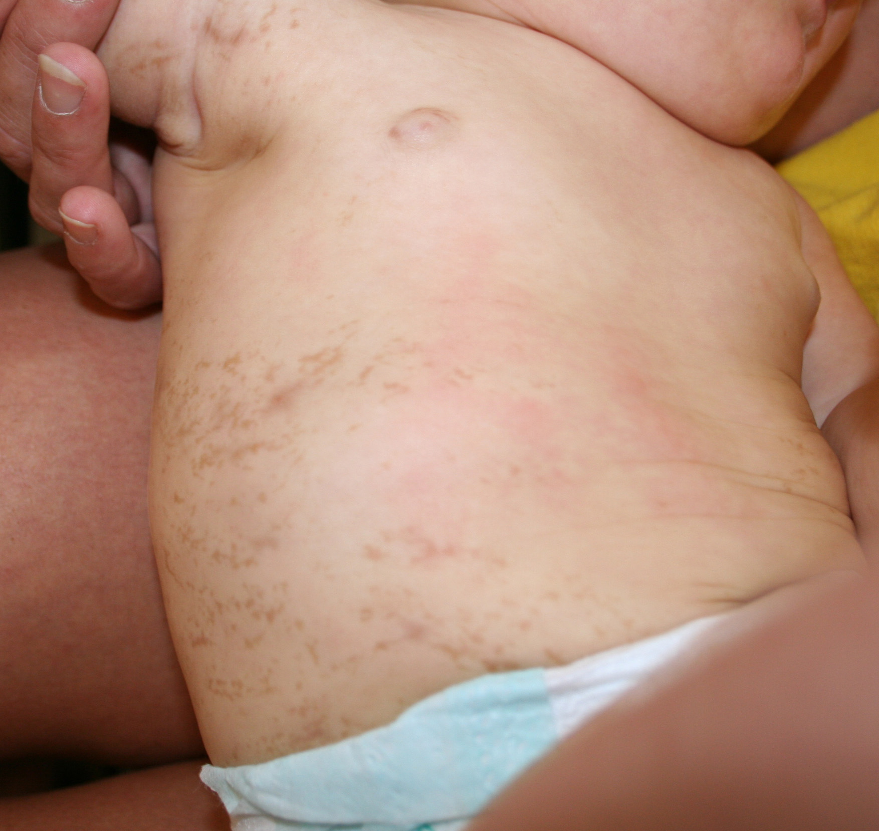
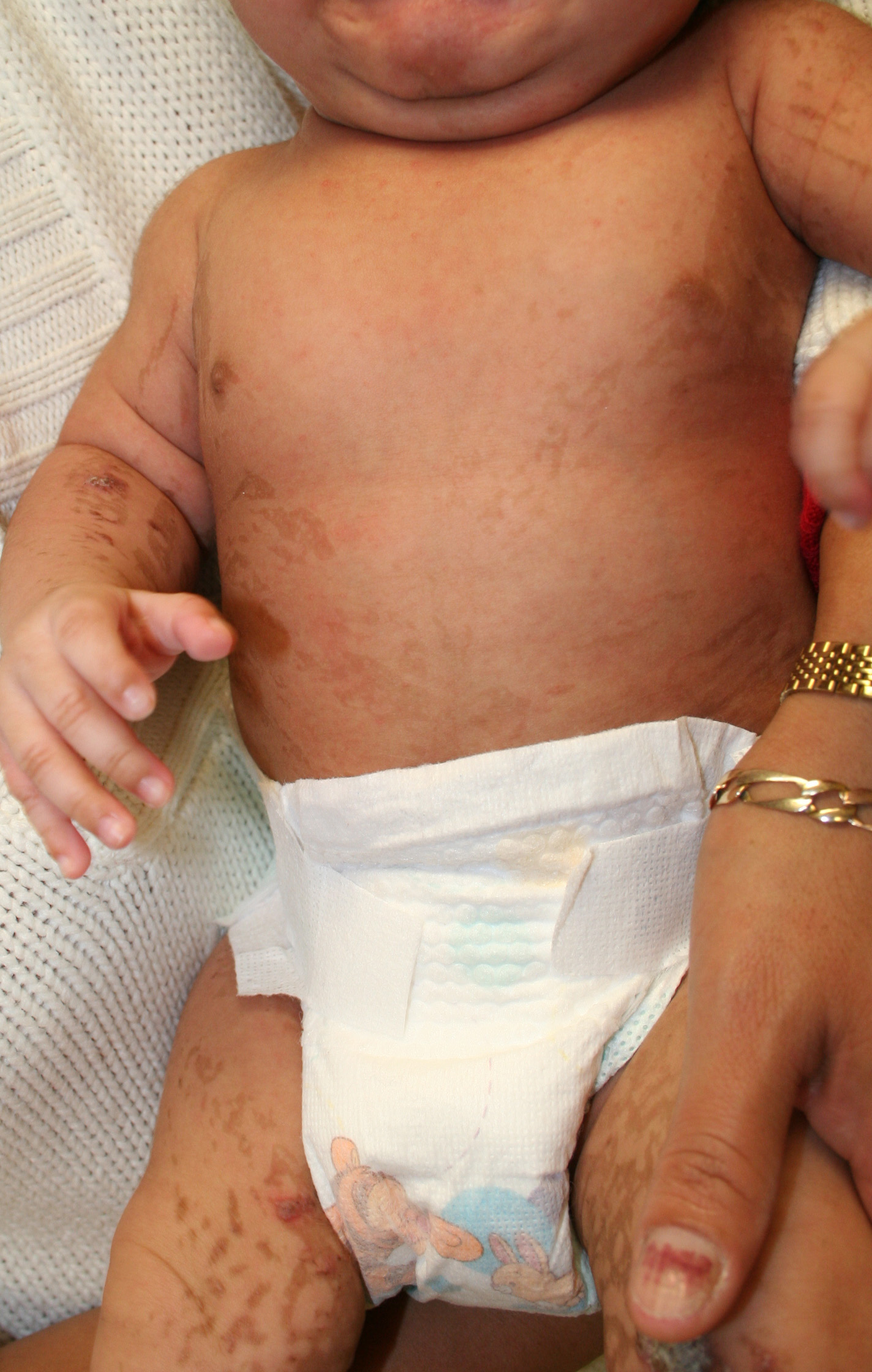
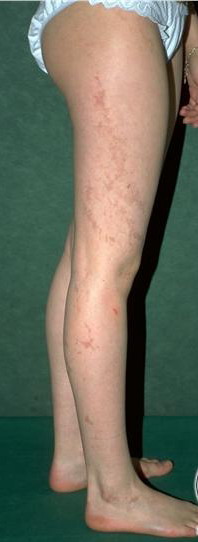
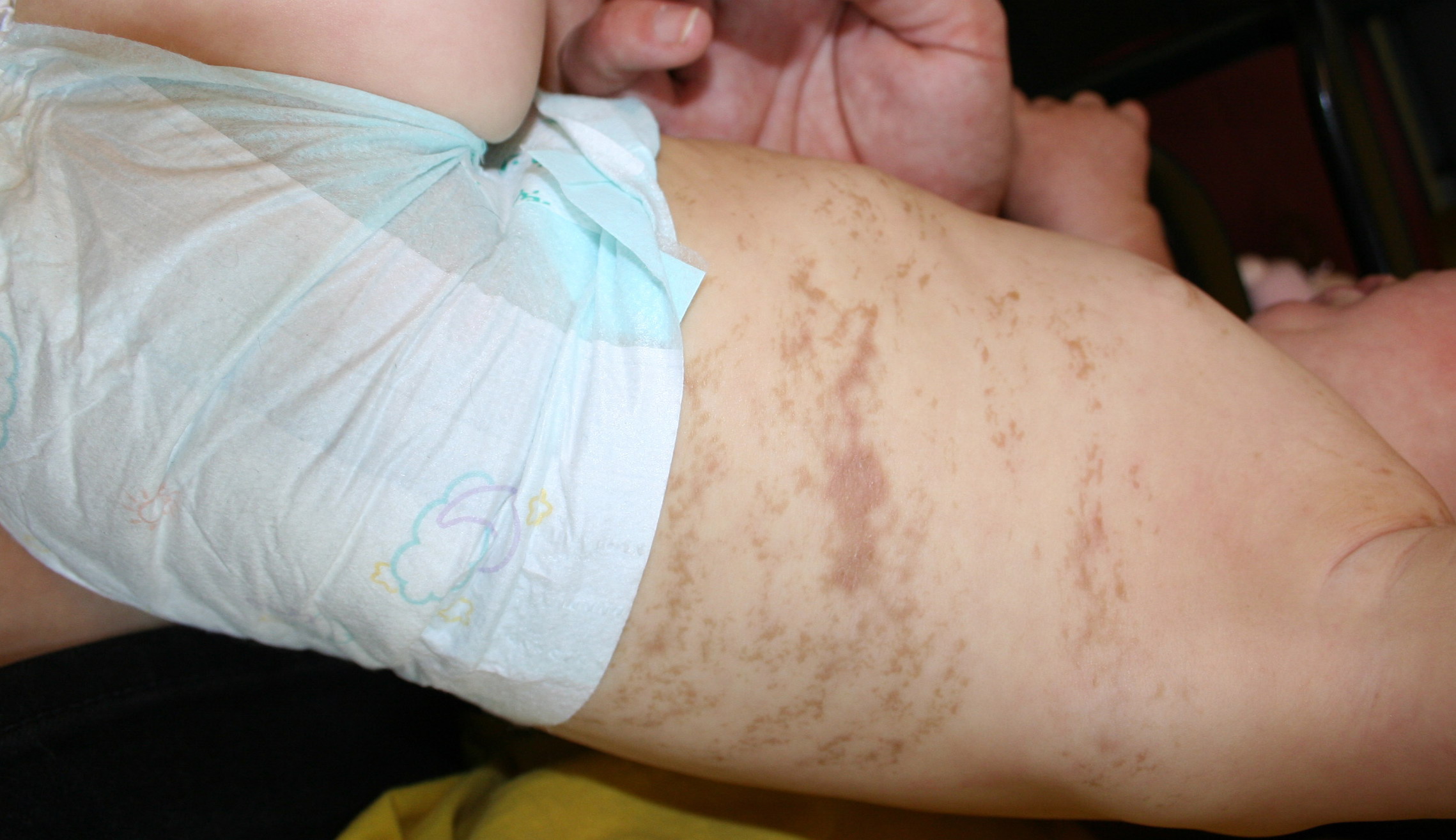
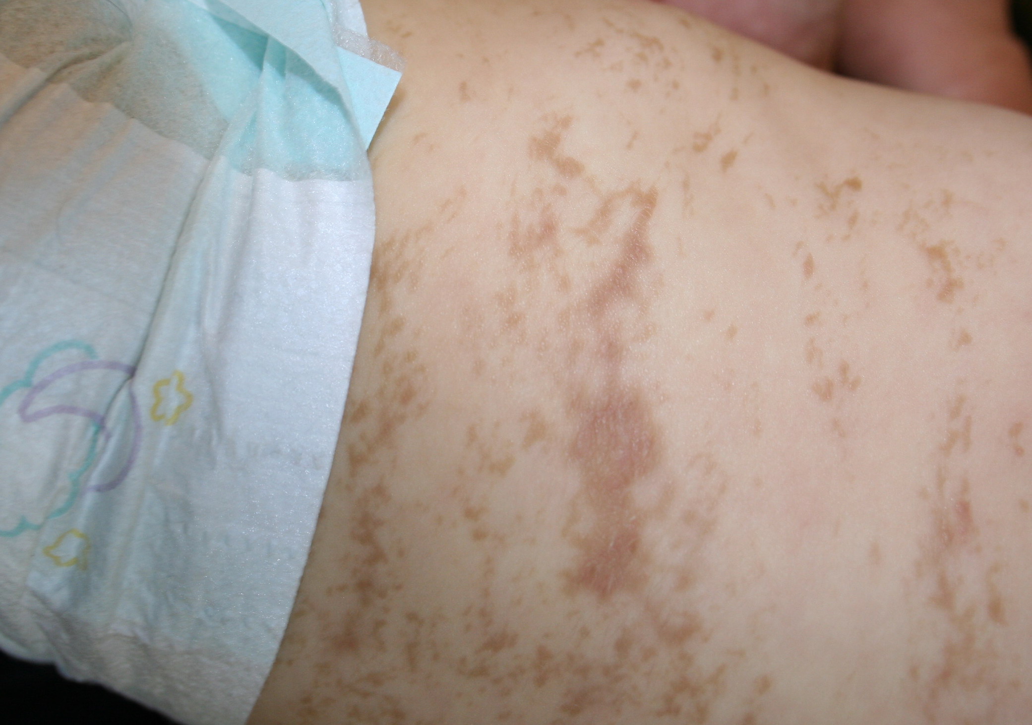
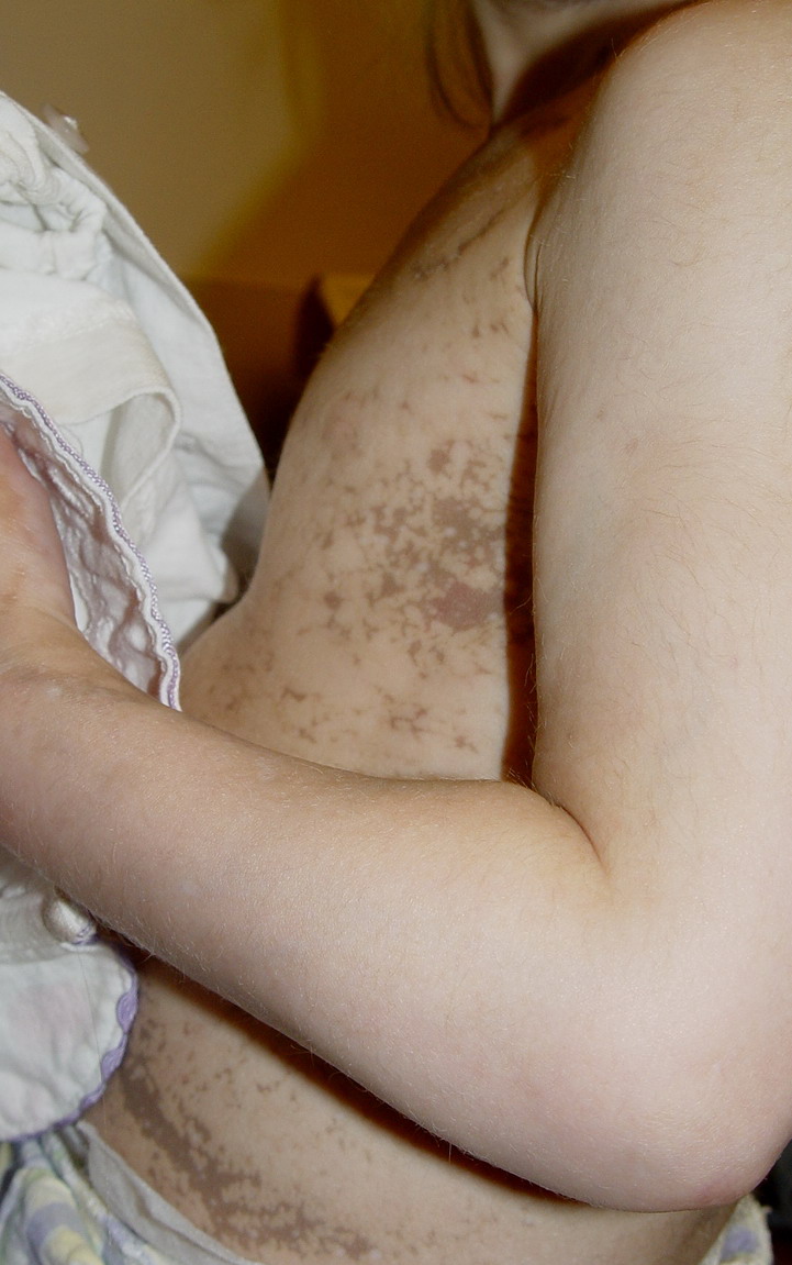
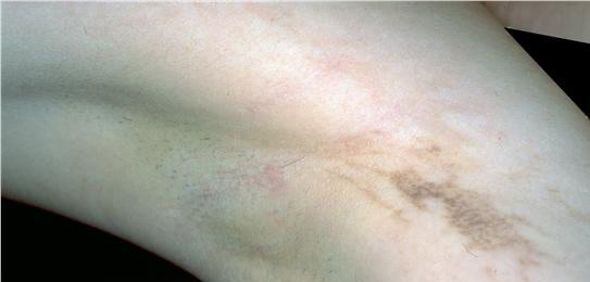
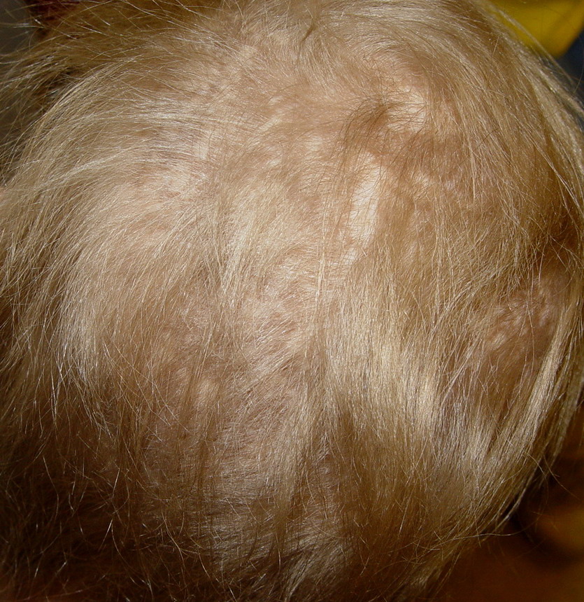
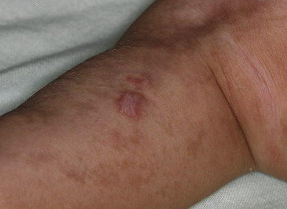
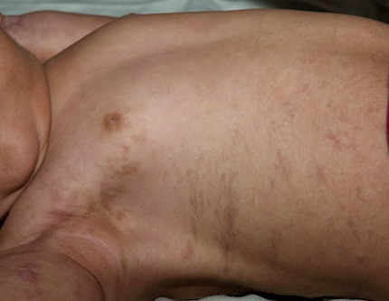
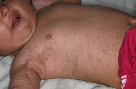
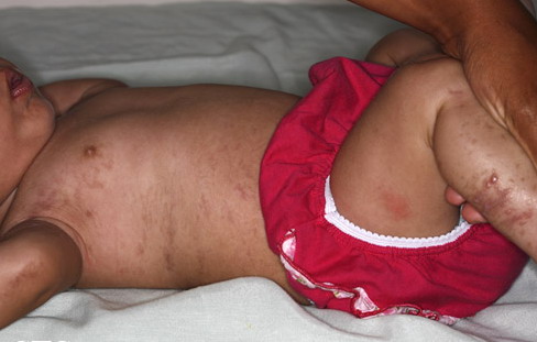
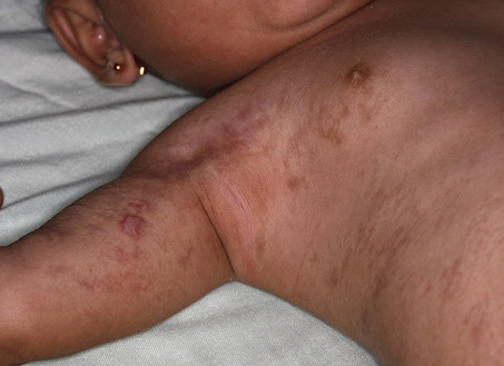
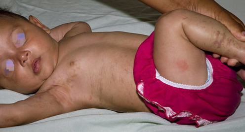
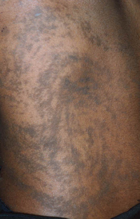
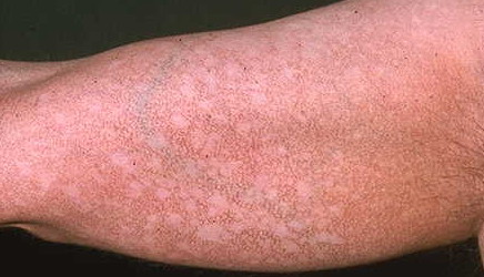
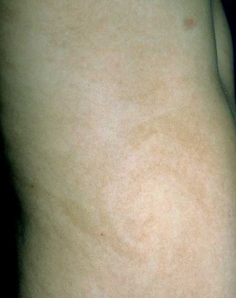
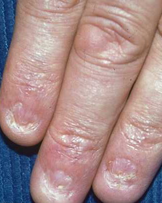
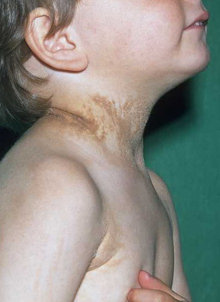
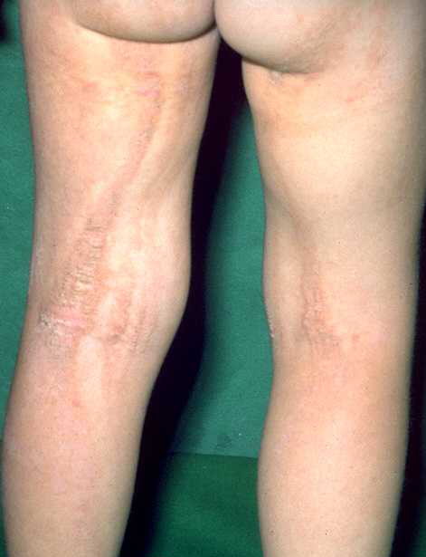
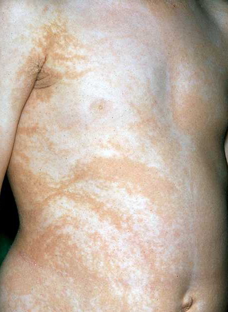
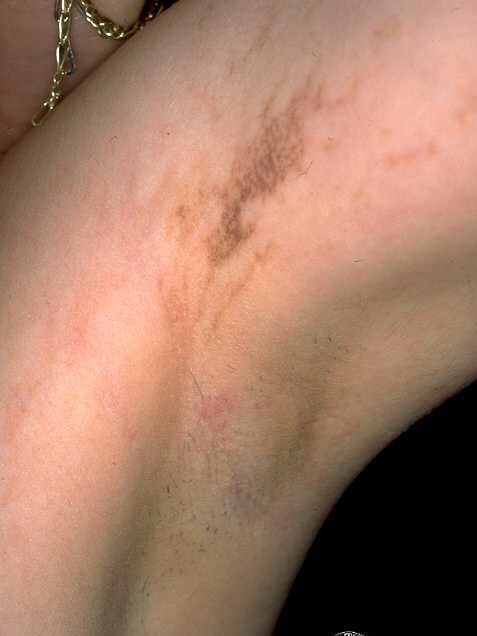
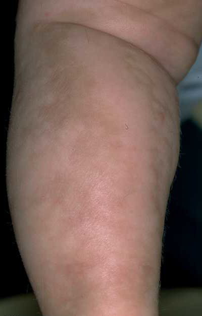
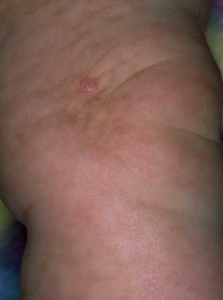
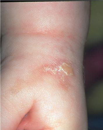
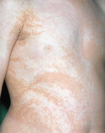
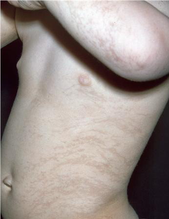
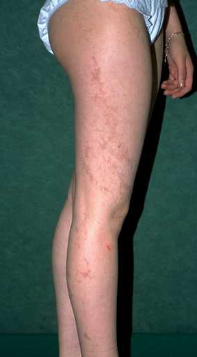
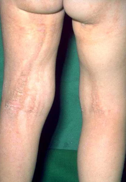
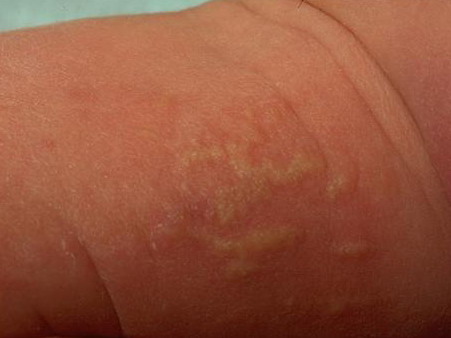
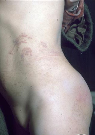
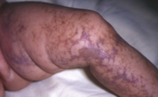
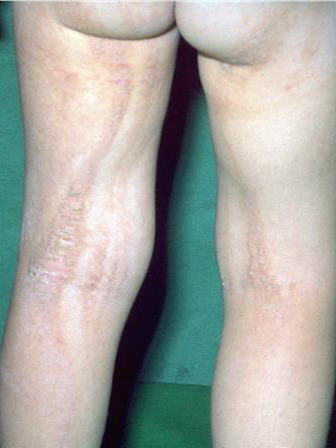
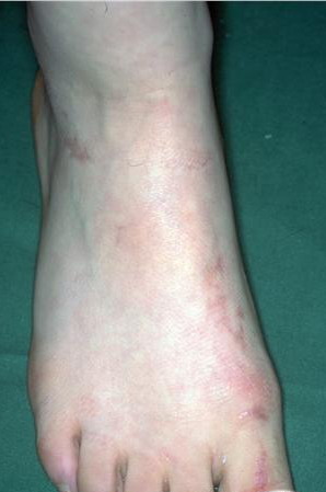
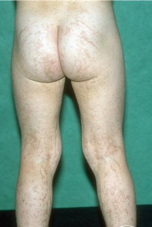
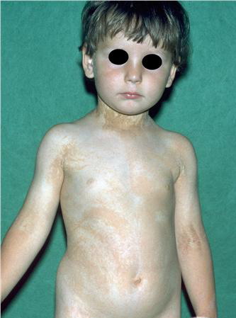
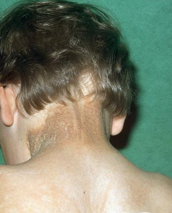
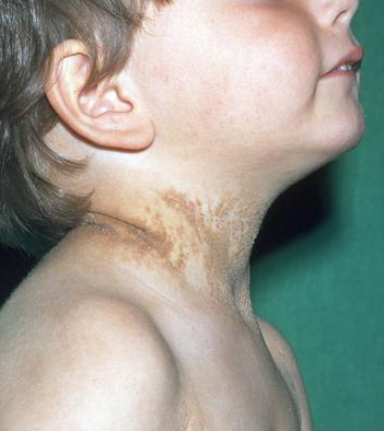
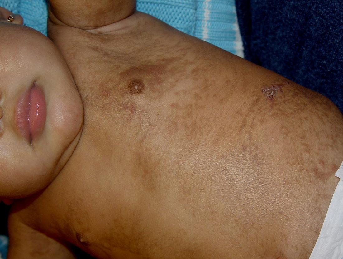
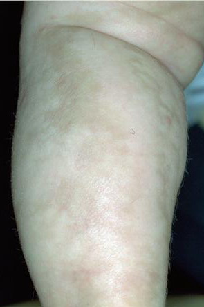
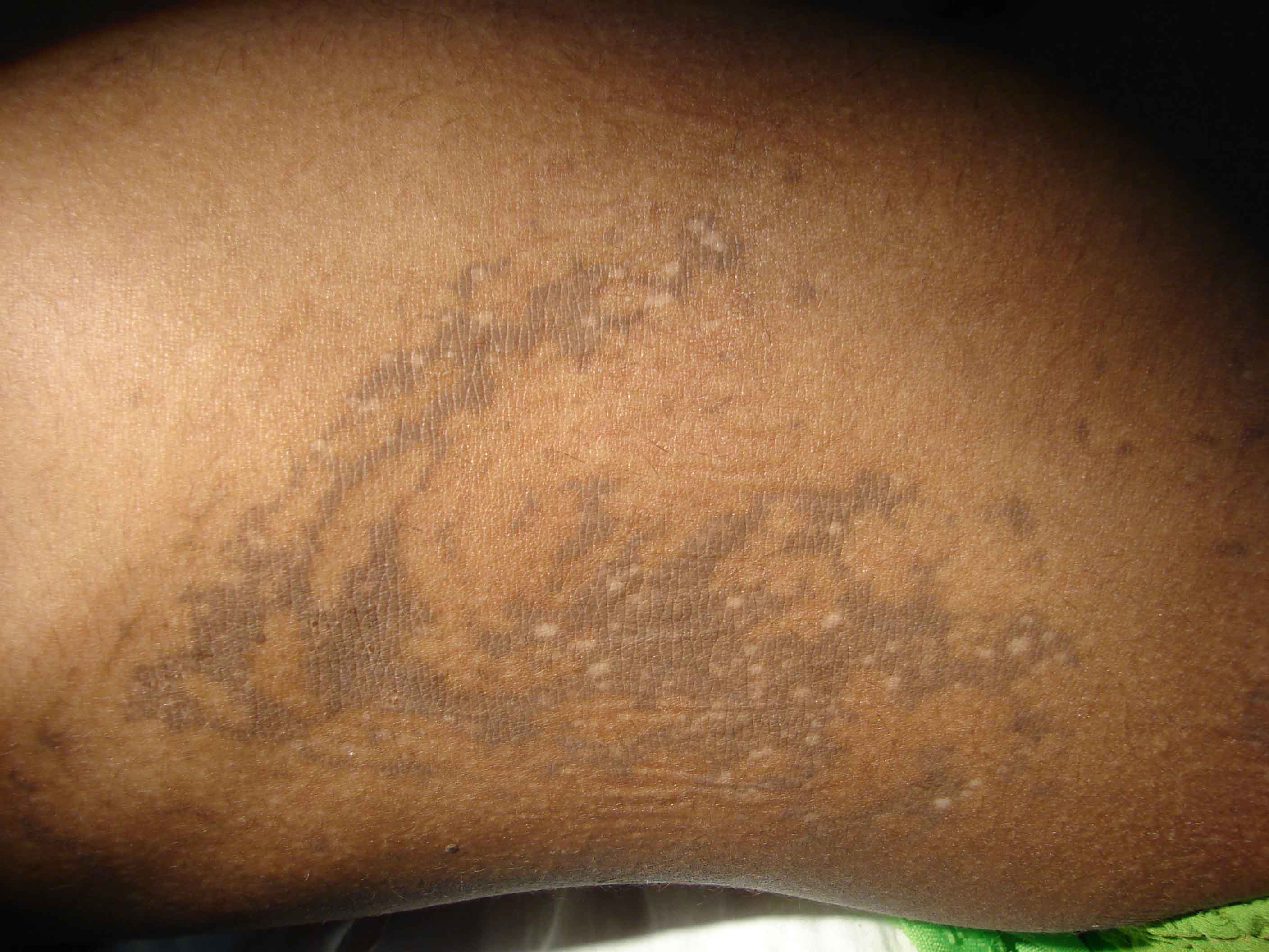
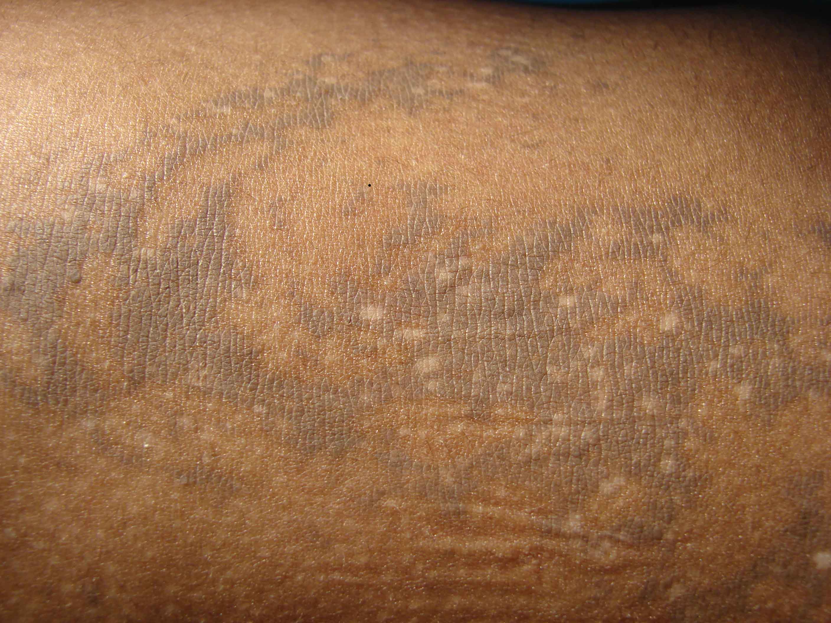
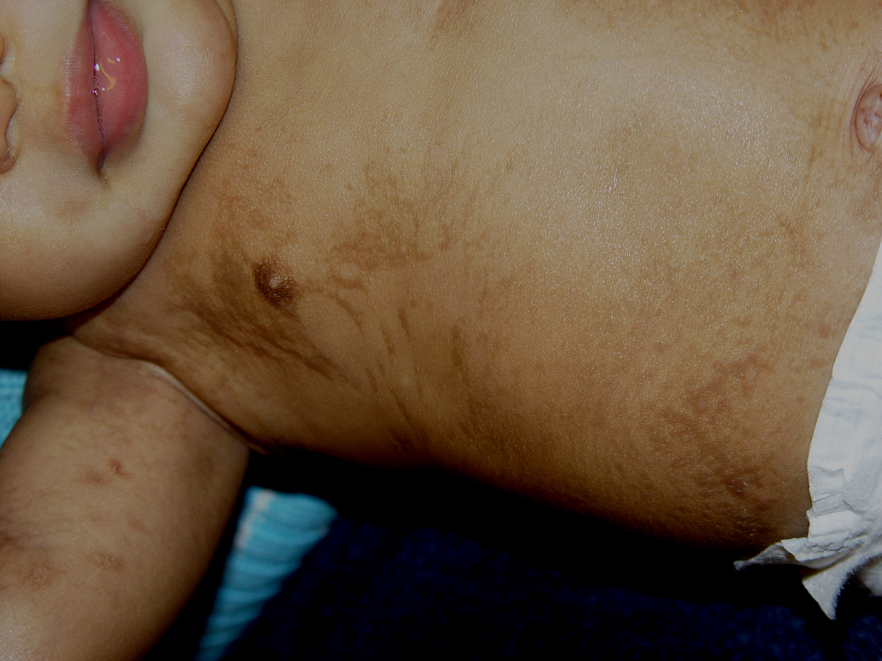
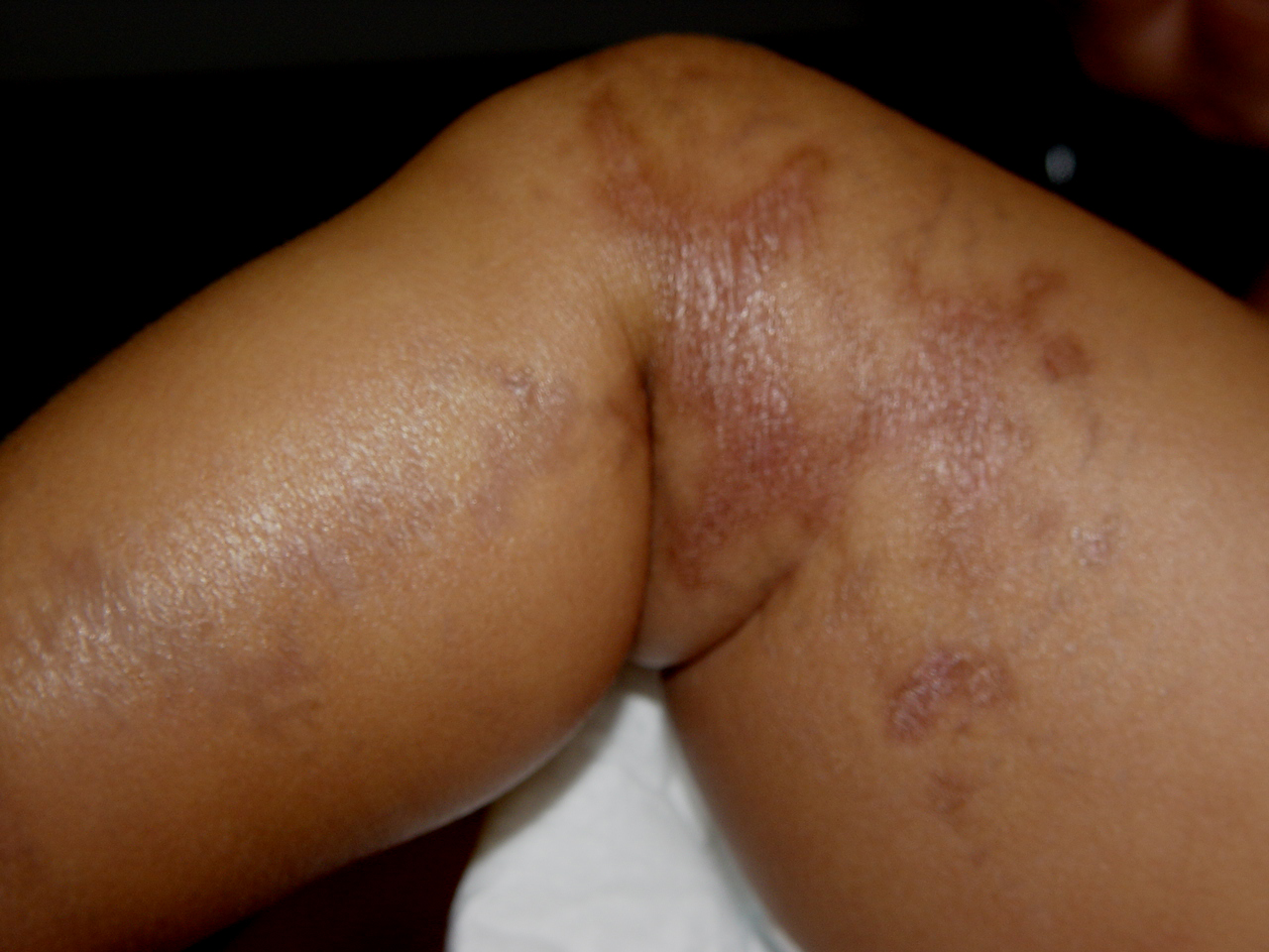
The hyperpigmentation (stage) appears in streaks and whorls along the lines of Blaschko and is usually most pronounced on the trunk, but they can also appear on the extremities. The degree of hyperpigmentation varies individually. Histologically, the areas of pigmentation show many melanin-laden melanophages, extensive deposits of melanin in the basal cell layer and dermis. There is vacuolization and degeneration in the epidermal basal cell layer. Usually, the hyperpigmentation fades gradually after several years and can become hypopigmented (stage 4), which represents post-inflammatory dermal scarring. The hypopigmentation stage is characterized by linear, atrophic, hairless scars following the Blaschko's lines.
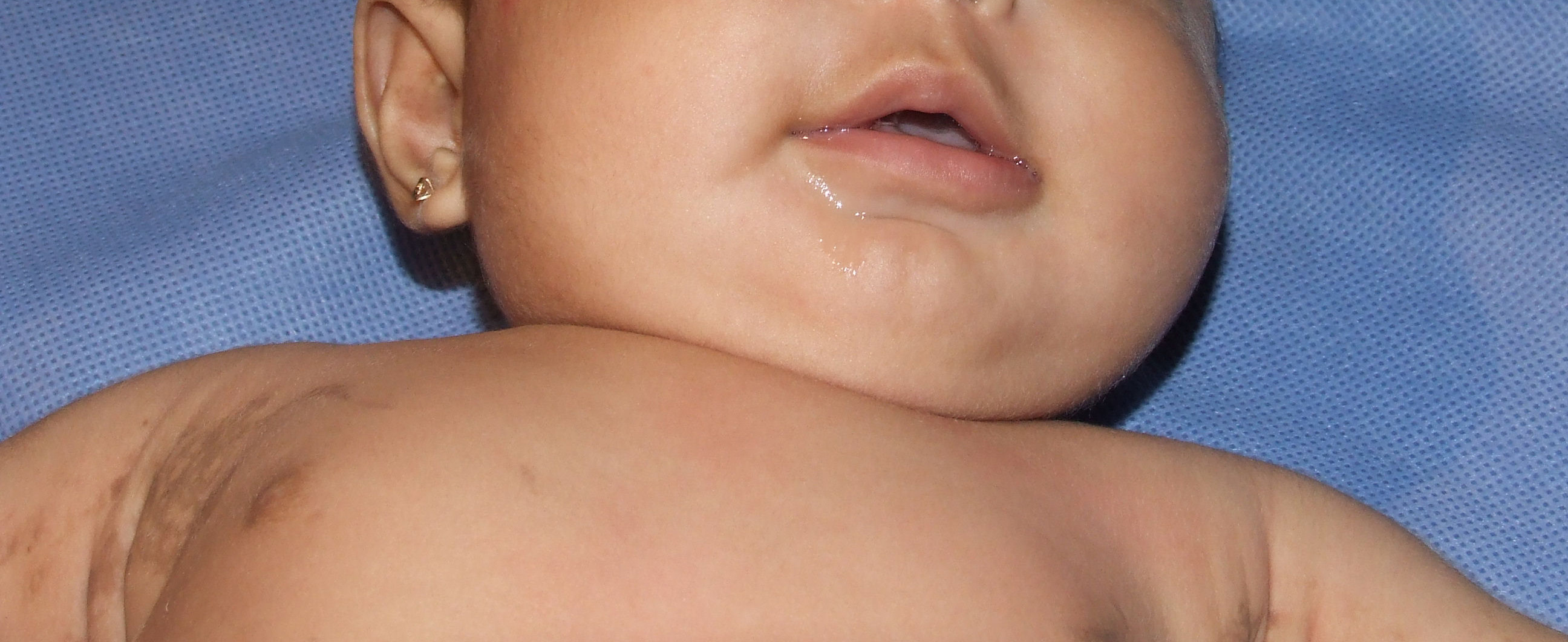
Histologically, the number of melanocytes seems to be normal, although a reduced number of melanocytes also has been reported. The epidermis is thinner and there is an absence or reduction of skin appendages in the dermis that may contribute to the impression of hypopigmentation.