|
MACULE
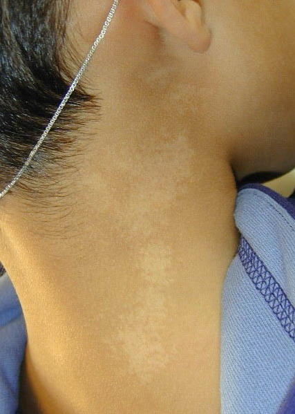
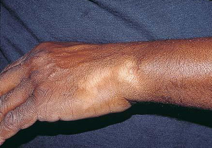
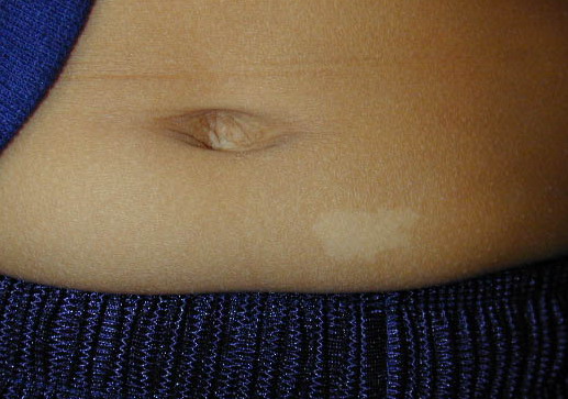
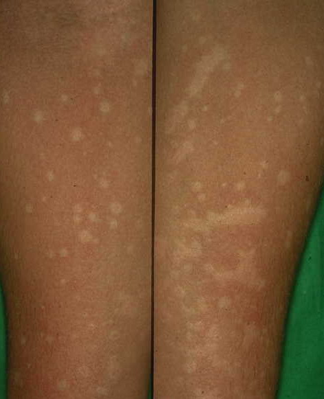
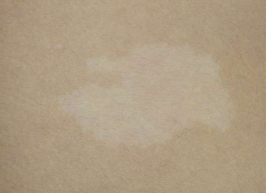
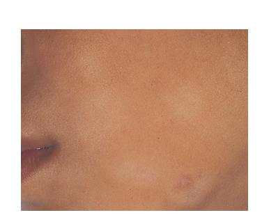
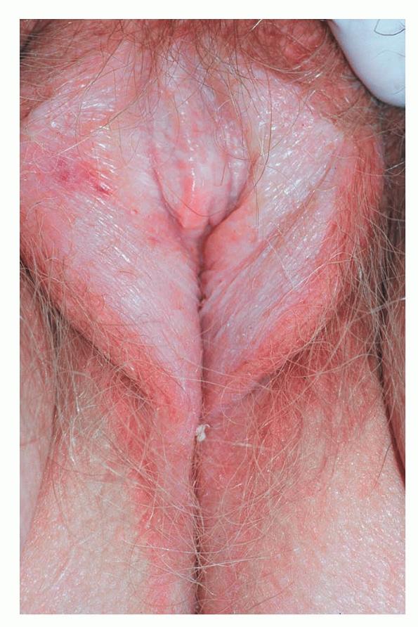
A macule is a flat lesion, even with the surface level of surrounding skin, perceptible as an area of color different from the surrounding skin or mucous membrane. Macules are non-palpable. Their shapes are varied and borders may be distinct or vague. Maculosquamous is a neologism invented to describe macules with fine non-palpable scaling, which may become apparent only after light scraping and scratching.
Perhaps the most important additional feature of a lesion other than primary morphology is color. Lesional color, which is often the first visual assessment made, is reliably reproducible with particular
types of pathologies, such as destruction of melanocytes, dilatation of dermal blood vessels, or inflammation of vessel walls with extravasation of red blood cells. As such, color provides meaningful insight into pathologic processes of the skin and facilitates clinical diagnosis. Pigmentary changes represent an important and common type of macular color change and may be described as hyperpigmented (as in post-inflammatory hyperpigmentation), hypopigmented (as in tinea versicolor), or depigmented (as in vitiligo).
|
Implications of Color Changes in Altered Skin
|
|
COLOR
|
PATHOLOGY
|
DIAGNOSTIC CONSIDERATION
|
|
Apple jelly
|
Granulomatous inflammation
|
Tuberculosis, sarcoidosis, leishmaniasis
|
|
Black
|
Melanin, necrosis
|
Melanoma, purpura fulminans, calciphylaxis
|
|
Blue
|
Deep dermal pigment, reduced hemoglobin, tattoo, medication
|
Blue nevus, amiodarone
|
|
Brown
|
Melanin, hemosiderin, chronic inflammation, post-inflammatory, dried serum
|
Nevus, melasma
|
|
Copper
|
Inflammation with plasma cells
|
Secondary syphilis
|
|
Green
|
Deep hemosiderin, pyocyanin pigment, tissue eosinophilia
|
Pseudomonas infection, tattoo, Wells syndrome
|
|
Grey
|
Deep melanin or other pigment deposition
|
Chloroquine toxicity, mongolian spot, erythema dyschromicum perstans
|
|
Lilac
|
Inflammation, dilatation of deep dermal blood vessels
|
Borders of evolving morphea, derma tomyositis
|
|
Orange
|
Granulomatous inflammation with histiocytes having abundant cytoplasm
|
Juvenile xanthogranuloma
|
|
Pearly
|
Epidermal proliferation without surface keratin
|
Basal cell carcinoma
|
|
Pink
|
Acute inflammation, dilatation of superficial dermal blood vessels, hemorrhage
|
Eczema
|
|
Red
|
Hemorrhage, acute inflammation, dilatation of blood vessels
|
Psoriasis, drug eruptions
|
|
Salmon pink
|
Inflammation with involvement of epidermis, dilatation of blood vessels inflammation with edema
|
Pityriasis rubra pilaris, psoriasis, urticaria
|
|
Violet
|
Hemorrhage, deep hemosiderin, lichenoid inflammation
|
Lichen planus, Kaposi sarcoma
|
|
White
|
Reduced or absent melanin synthesis, post-inflammatory
|
Tinea versicolor, albinism, vitiligo
|
|
Yellow
|
Superficial staphylococcus or streptococcus infection mixed with keratinized cells, carotenoids, hemosiderin, bile pigment, accumulated lipid
|
Impetigo, xanthomas, sebaceous hyperplasia, necrobiosis lipoidica diabeticorum, jaundice
|
|
|

