▪ EPIDERMOLYTIC
HYPERKERATOSIS
In 1902, Brocq described bullous ichthyotic erythroderma and distinguished the blistering type from the nonblistering type of congenital ichthyotic erythroderma. The original description included three unrelated patients whose clinical manifestations varied. However, this was probably the first description of
epidermolytic hyperkeratosis . Today, the disease is named for the distinctive histopathologic features of vacuolar degeneration of the epidermis (i.e., epidermal lysis) and associated hyperkeratosis. Epidermolytic hyperkeratosis is also known as bullous congenital ichthyosiform erythroderma, an earlier descriptive name signifying the blistering, neonatal presentation, scaling, and redness.
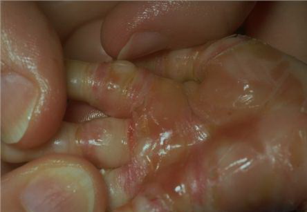
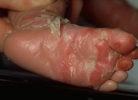
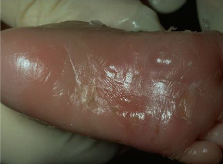
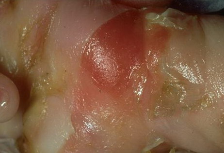
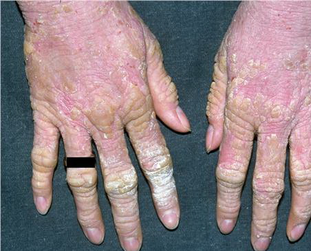
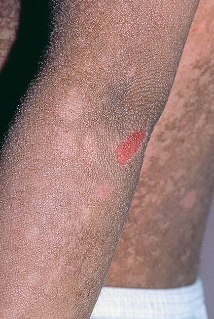
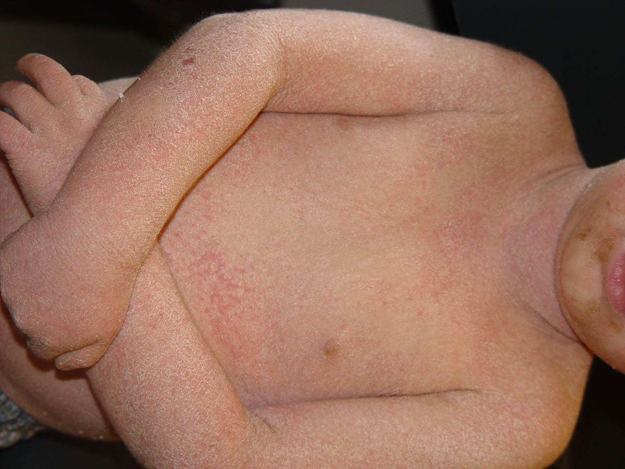
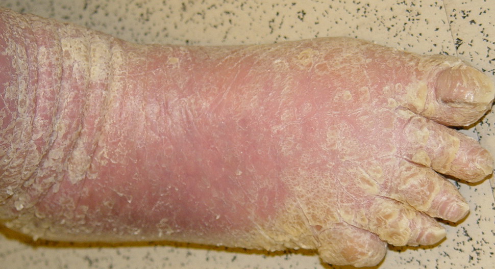
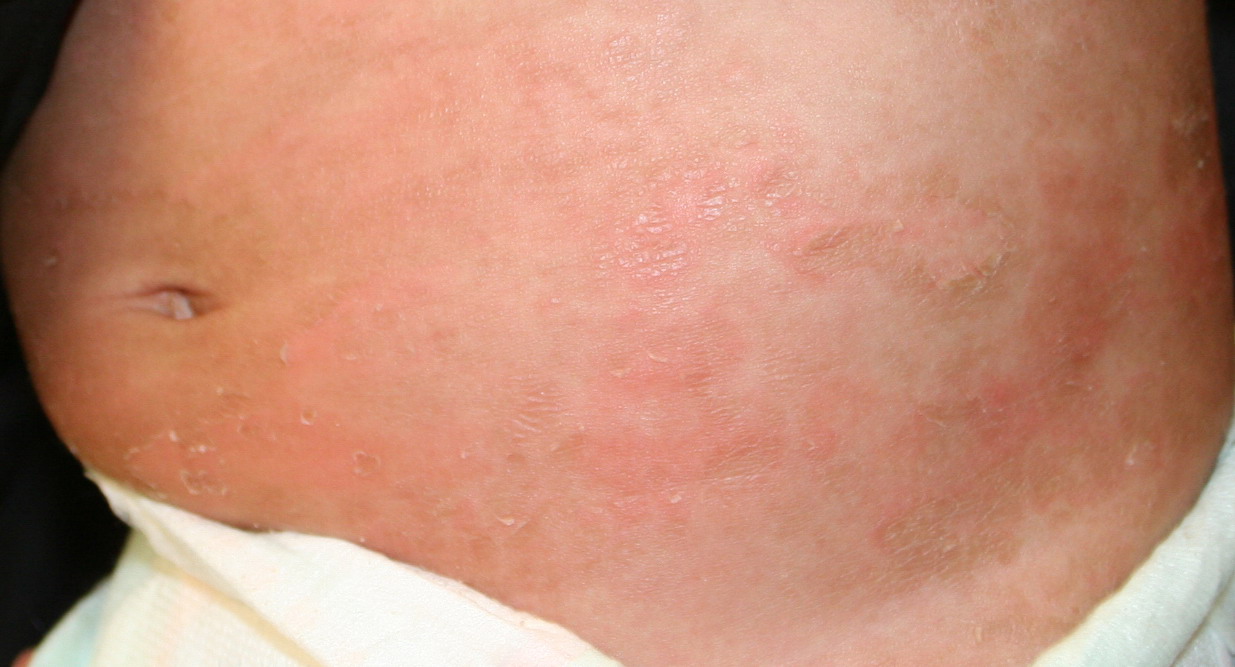
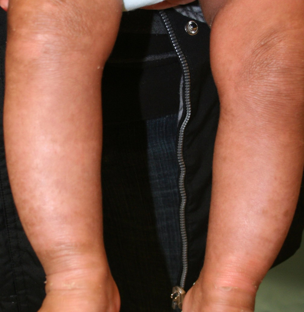
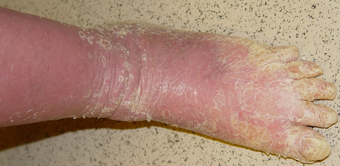
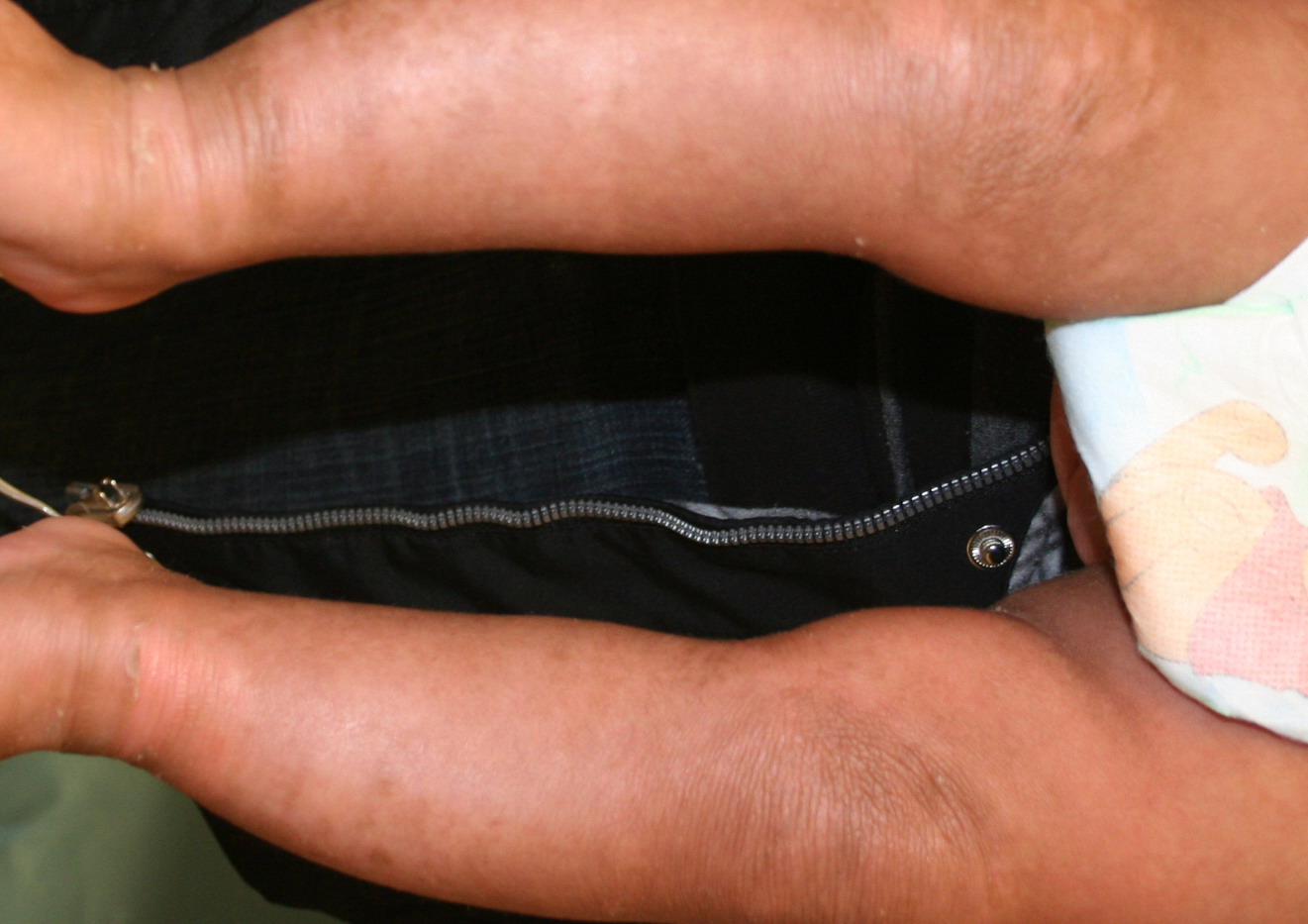
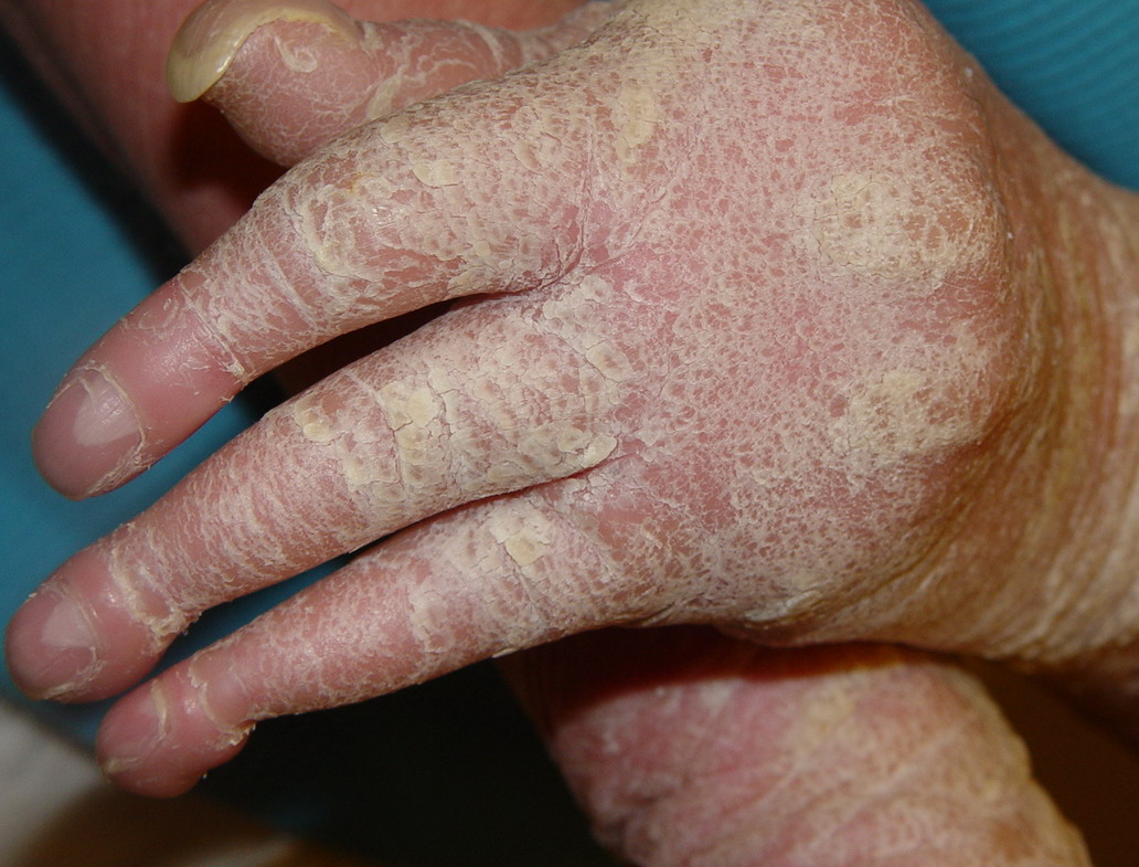
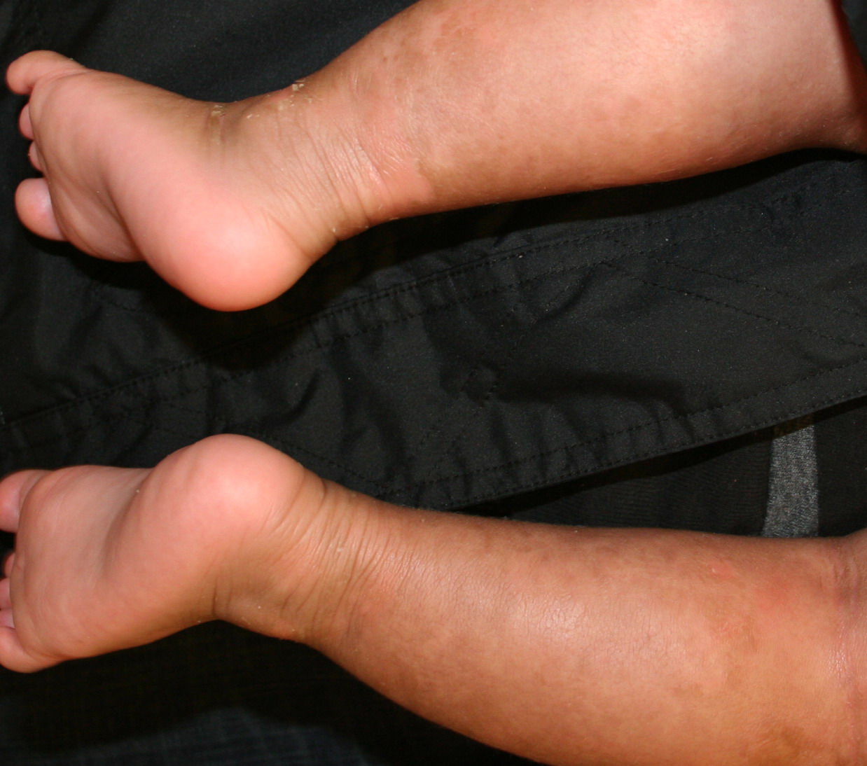
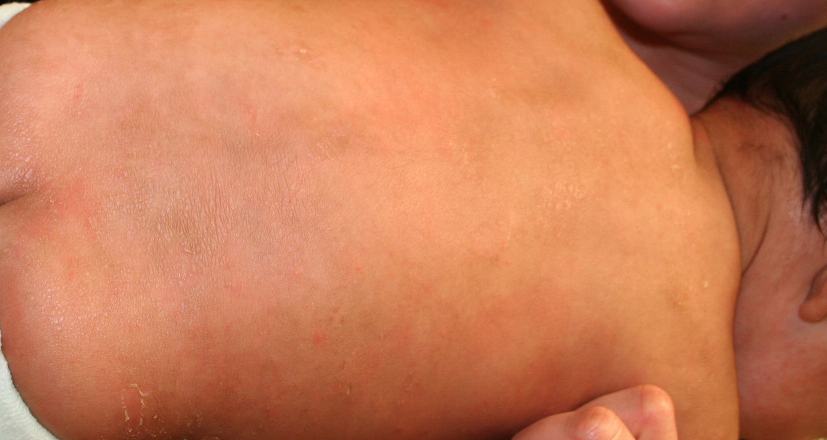
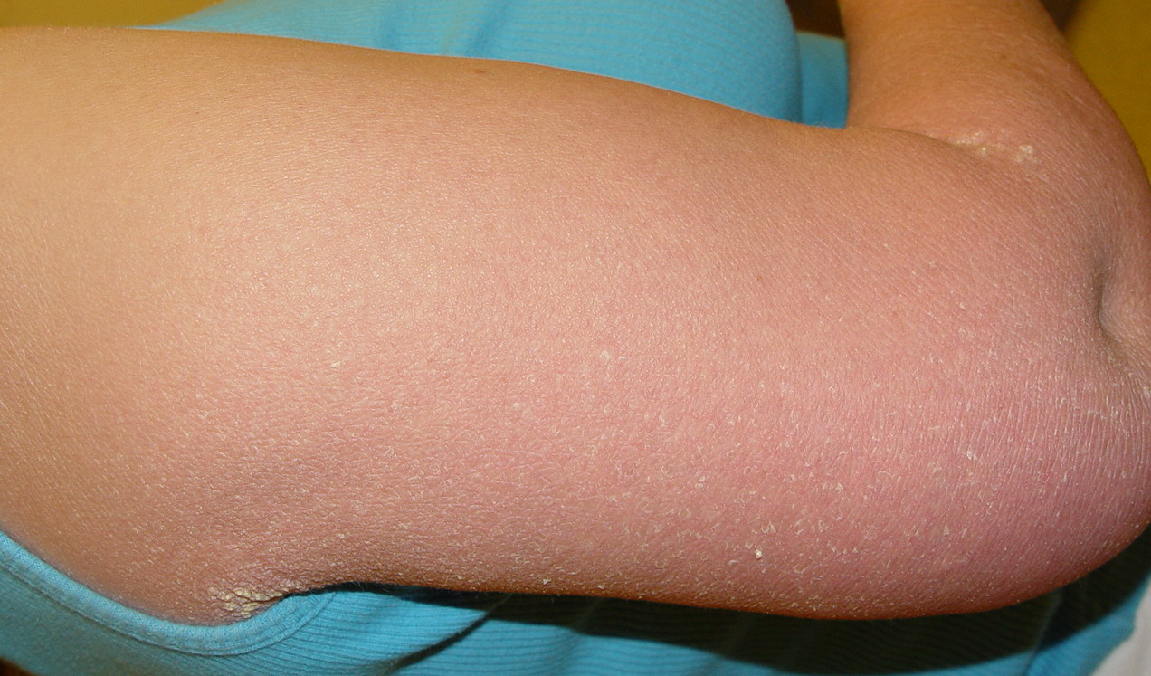
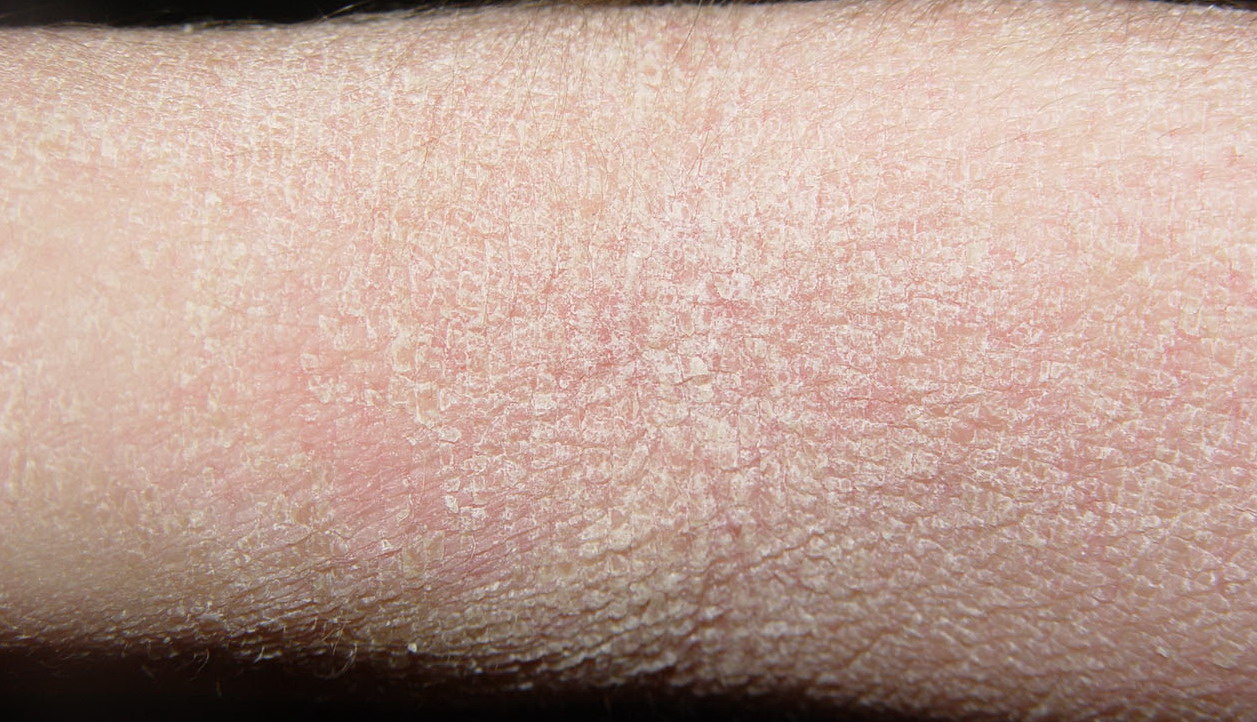
Epidermolytic hyperkeratosis is transmitted as an autosomal dominant trait with a prevalence of approximately 1 in 200,000 to 300,000 persons. However, there is a high frequency of spontaneous mutation, and as many as one-half of the cases have no family history and represent new mutational events. The disease usually presents at birth with blistering, redness, and peeling . With time, generalized hyperkeratosis may develop, which may or may not be associated with erythroderma. Epidermolytic hyperkeratosis skin usually has a characteristic pungent odor, thought to be related to superinfection by mixed flora.
|
Characteristics of Epidermolytic Hyperkeratosis
|
|
|
NPS-1
|
NPS-2
|
NPS-3
|
PS-1
|
PS-2
|
PS-3
|
|
Palm/sole hyperkeratosis
|
-
|
-
|
-
|
+
|
+
|
+
|
|
Palm/sole surface
|
Normal
|
Normal
|
Hyperlinear, minimal scale
|
Smooth
|
Smooth
|
Cerebriform
|
|
Digital contractures
|
-
|
-
|
-
|
-
|
+
|
-
|
|
Scale
|
Hystrix
|
Brown
|
Fine, white
|
Mild
|
White scale to peel
|
Tan
|
|
Distribution
|
Generalized
|
Generalized
|
Generalized
|
Localized
|
Generalized
|
Generalized
|
|
Erythroderma
|
-
|
-
|
+
|
-
|
+
|
-
|
|
Blistering
|
+
|
+
|
+
|
Localized
|
+
|
Neonatal
|
|
Most common mutation
|
Keratin-10
|
Keratin-1
|
|
NPS = types without severe palm/sole hyperkeratosis; PS = types with severe palm/sole hyperkeratosis; - = absent; + = present.
|
|
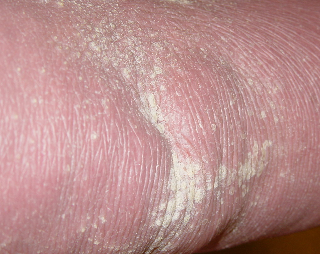
In contrast to most other ichthyoses, the histopathologic picture of epidermolytic hyperkeratosis is distinctive. There are a tremendously thickened stratum corneum and vacuolar degeneration of the upper epidermis, leading to the histologic term epidermolytic hyperkeratosis . The vacuolar degeneration usually involves the upper epidermis and occasionally all of the suprabasilar keratinocytes. Granular cells exhibit dense, enlarged, irregularly shaped masses that appear to be keratohyalin granules. On electron-microscopic examination, clumping of filaments is observed to begin in the first suprabasal layer. These aggregated filaments are clumps of keratin intermediate filaments that contain the terminal differentiation-specific keratin-1 and keratin-10.
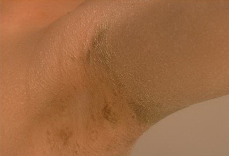
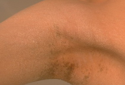
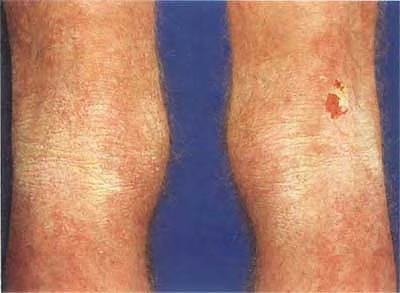
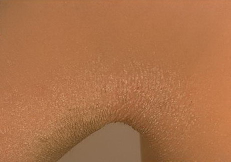
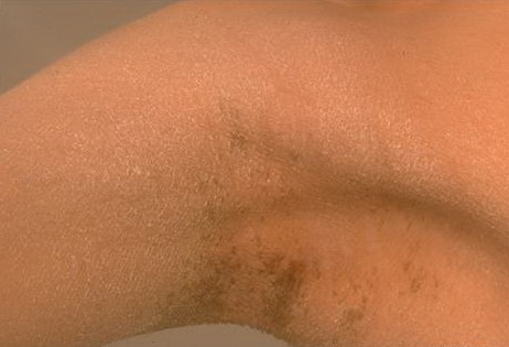
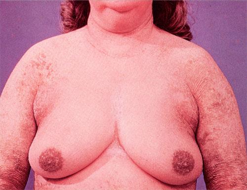
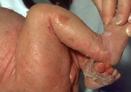
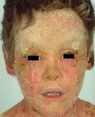
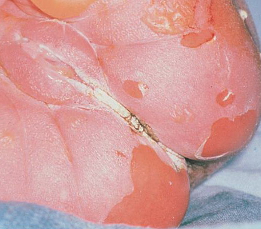
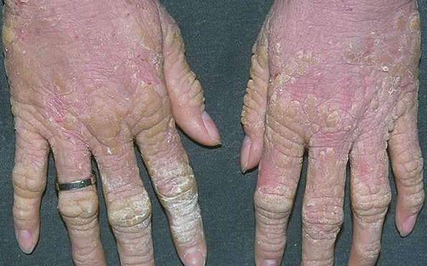
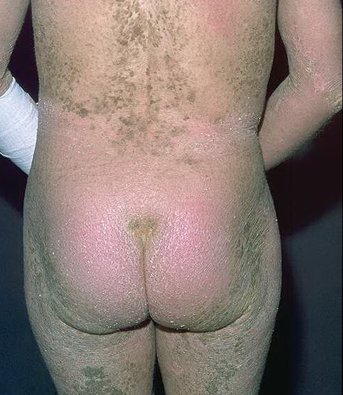
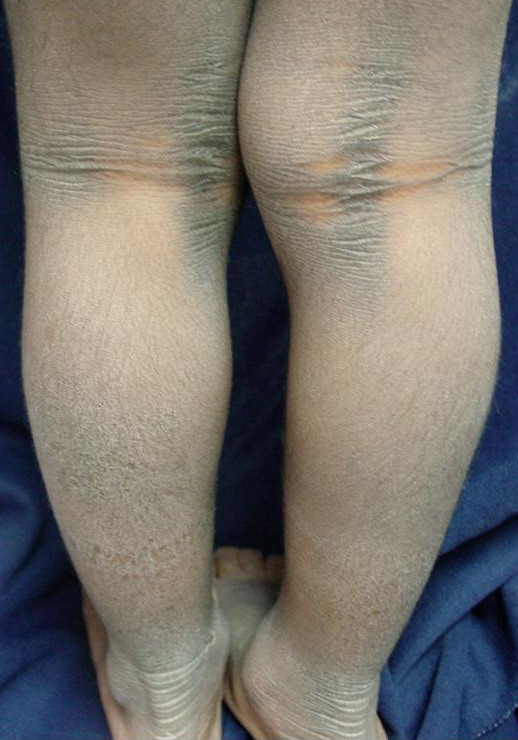
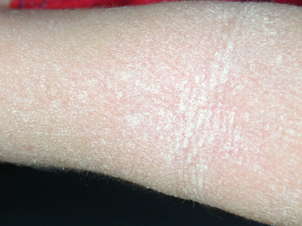
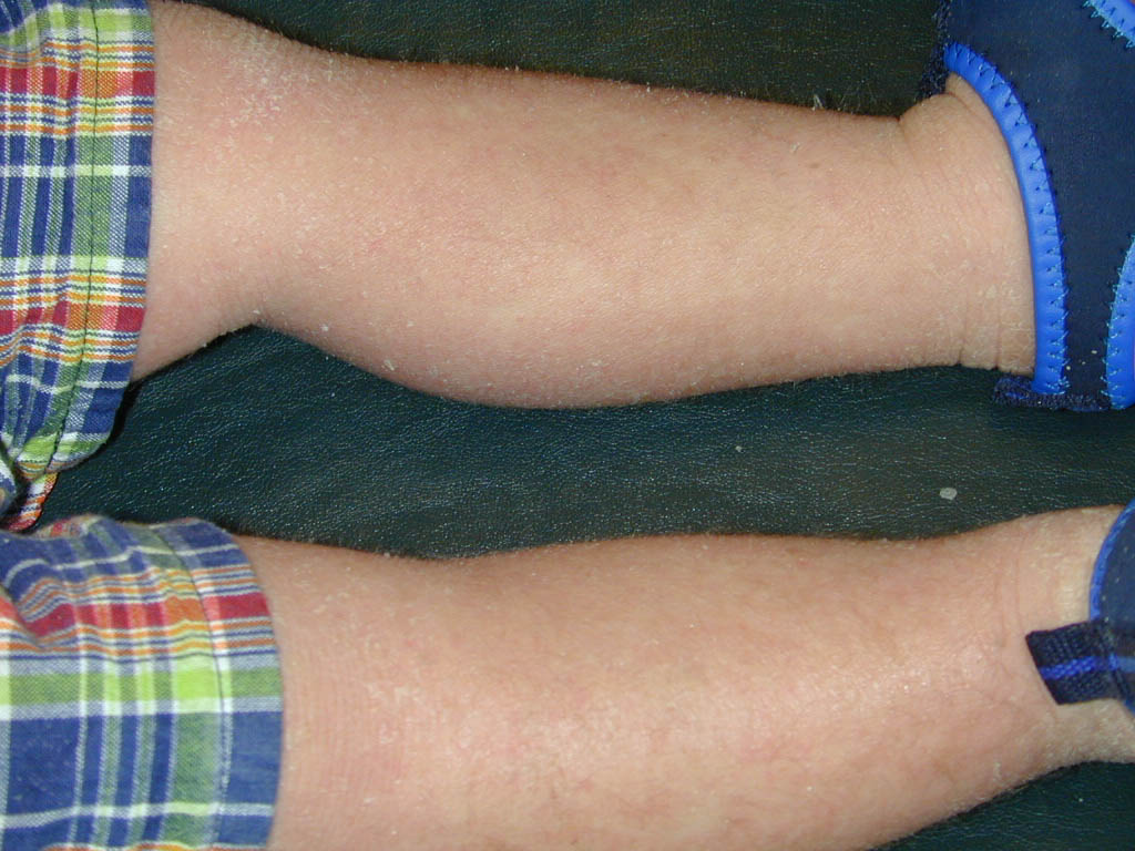
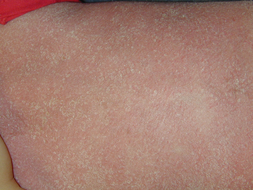
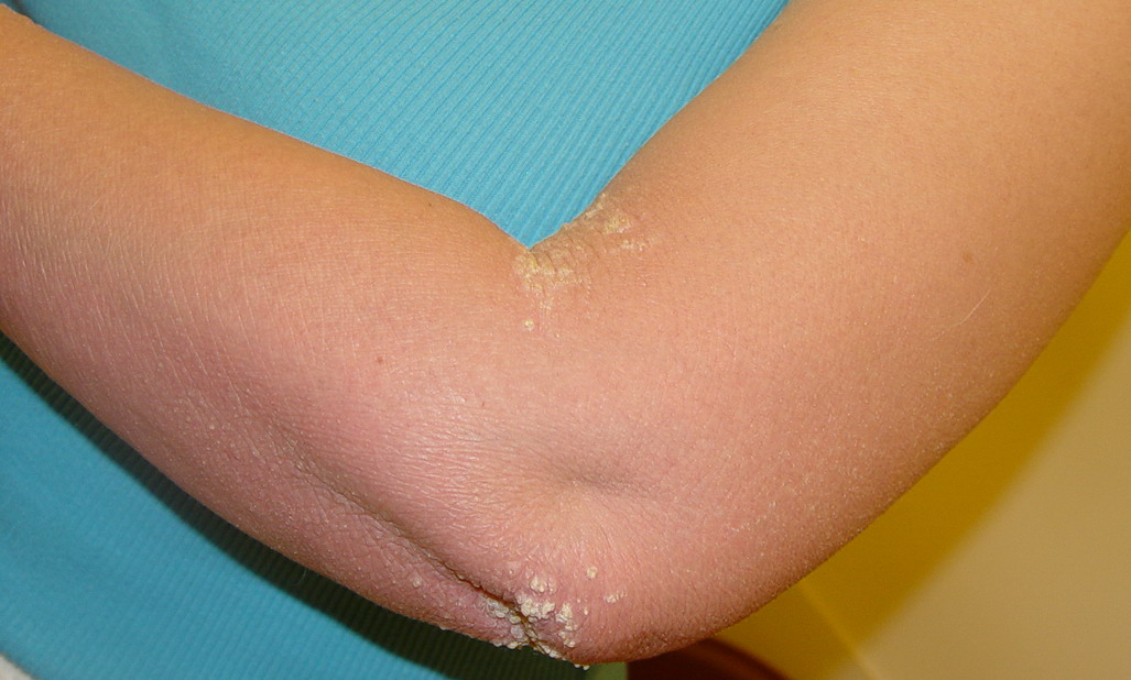
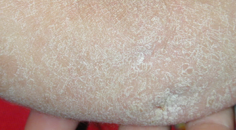
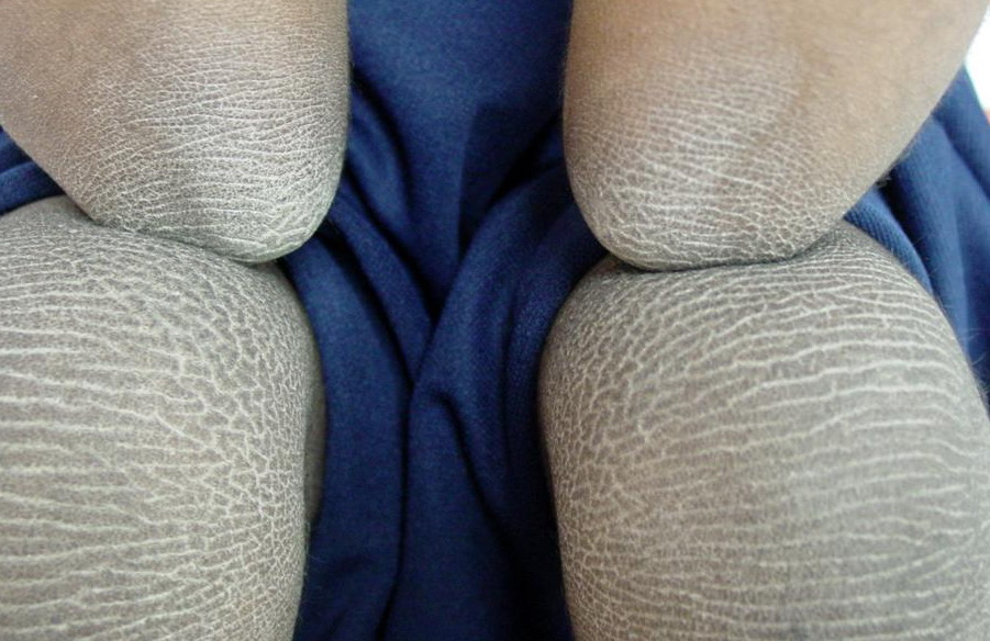
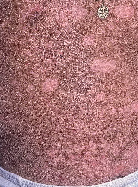
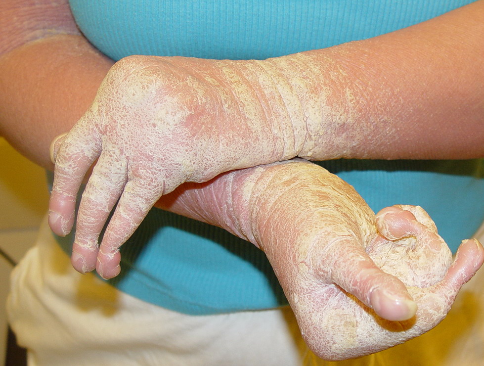
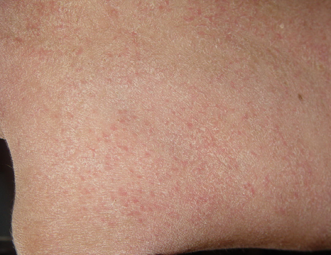
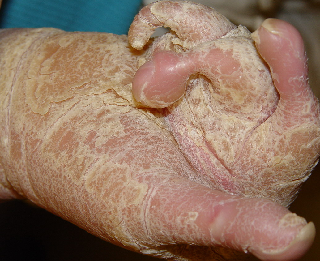
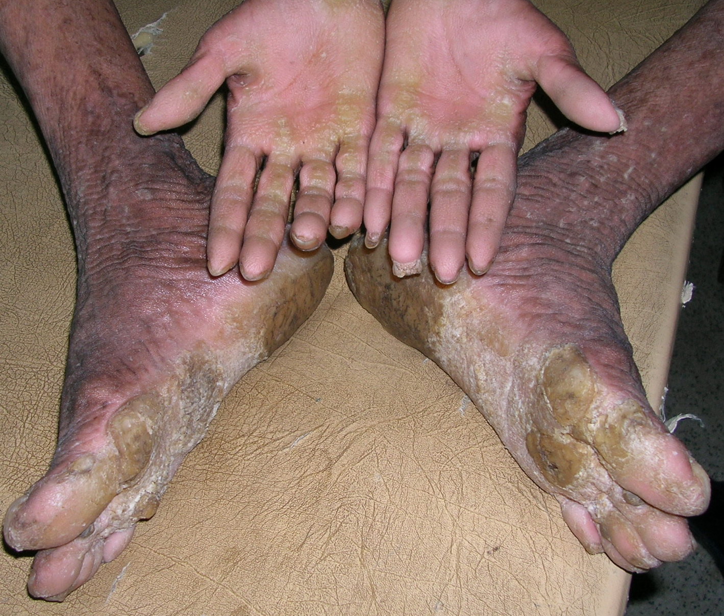
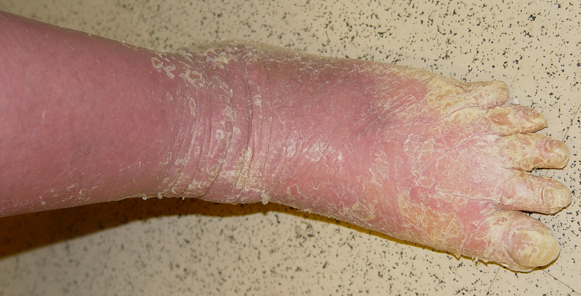
There is striking clinical heterogeneity between epidermolytic hyperkeratosis families. Six distinctive clinical phenotypes have been described. Several clinical features can be useful to distinguish between the different sub-types, including the presence versus absence of severe palmar/plantar hyperkeratosis and of erythroderma, quality of scale, extent of involvement, presence of digital contractures, and gait abnormality . Three types with no severe palm/sole hyperkeratosis are called NPS types. Patients with the NPS-1 clinical phenotype have generalized involvement with thick, brown verrucous hyperkeratosis, most prominent over joints. Palms and soles are spared. Cobblestone or hystrix (porcupine-like) spiny papules are characteristic; this severe clinical presentation is one of the more commonly recognized types of epidermolytic hyperkeratosis . NPS-2 is similar, but much milder, without the hystrix spines and with relative sparing of the skin between joints . Generalized exfoliative erythroderma is found in NPS-3 . Three types with severe palm/sole hyperkeratosis are called PS types. Patients with the PS-1 type have a smooth palmar/plantar hyperkeratosis with a sharp border, often delineated by a red halo. Blisters may be present at the border. There may be limited involvement of the trunk, usually confined to areas over joints . The PS-2 type is characterized by generalized erythroderma and scaling. Volar involvement may be severe, with contractures of the digits and ainhum-like constricting digital bands . The PS-3 type has generalized skin involvement with a pebbly hyperkeratosis that is arrayed in a distinctive, cerebriform pattern on the palms and soles .
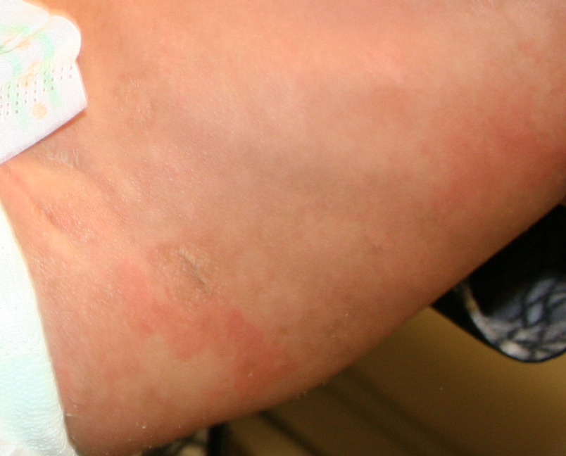
Sporadic epidermolytic hyperkeratosis due to a postzygotic, spontaneous mutation during embryogenesis can present in a mosaic pattern of skin involvement. Areas of hyperkeratosis alternating with normal skin are often distributed in streaks along Blaschko's lines . These may be limited to a few streaks, or there may be many, with widespread, patchy involvement. Unilateral localization can also occur. This clinical mosaic pattern correlates with underlying genetic mosaicism in that keratin mutations characteristic of epidermolytic hyperkeratosis, which were found in lesional skin, were absent in normal skin.58,59 If the germline is involved, individuals with mosaic epidermolytic hyperkeratosis can transmit the mutation, which leads to generalized epidermolytic hyperkeratosis in affected offspring
Within keratinocytes, keratin intermediate filaments form an elaborate network that confers structural stability to the cells. In the suprabasilar differentiating keratinocytes of interfollicular epidermis, this network is formed by keratin-1 and keratin-10. In epidermolytic hyperkeratosis, failure of this network leads to keratinocyte fragility (particularly of the upper epidermis), easy blistering, abnormal epidermal kinetics (hyperproliferation), and thickened stratum corneum (hyperkeratosis). Keratin-10 is the co-expressed partner of keratin-1, both of which are required to form keratin intermediate filaments in the cells of the suprabasal layers of the epidermis . Genetic linkage studies and sequencing of the keratin-1 and keratin-10 genes of a number of epidermolytic hyperkeratosis families have identified mutations in either keratin-1 or keratin-10. In most cases, palmar plantar involvement implies mutations in keratin-1; this may reflect the “redundancy” of keratin-9 (a keratin that occurs only in the suprabasal epidermis of palmar and plantar skin) and keratin-10 in palmar/plantar epithelium. Mutations in keratin-9 have been found in families with the type of epidermolytic hyperkeratosis limited exclusively to the palms and soles (Vörner)