|
Elastosis-a form of solar aging = المران كشكل من شيخوخة الجلدالضيائية |
 |
 |
 |
|
Features of Photoaged Skina
|
|
CLINICAL
|
HISTOLOGIC
|
|
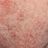
Dryness (roughness)
|
Increased compaction of stratum corneum, increased thickness of granular cell layer, reduced epidermal thickness, reduced epidermal mucin content
|
|
Actinic keratoses
|
Nuclear atypia, loss of orderly, progressive keratinocyte maturation; irregular epidermal hyperplasia and/or hypoplasia; occasional dermal inflammation
|
|
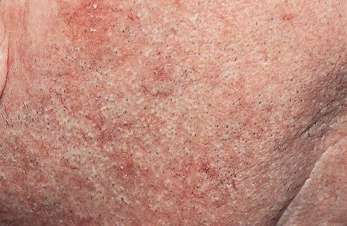
Irregular pigmentation
|
|
|
|
Freckling
|
Reduced or increased number of hypertrophic, strongly DOPA-positive melanocytes
|
|
|
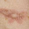
Lentigines
|
Elongation of epidermal rete ridges; increases in number and melanization of melanocytes
|
|
|
Guttate hypomelanosis
|
Reduced number of atypical melanocytes
|
|
|
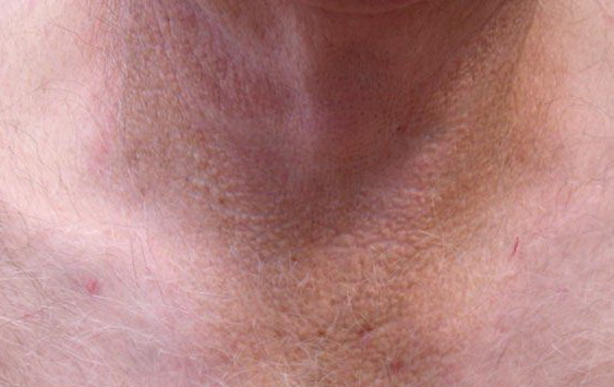
Diffuse irreversible hyperpigmentation
|
Increased number of DOPA-positive melanocytes and increased melanin content per unit area and increased number of dermal melanophages
|
|
Wrinkling
|
|
|
|
Fine surface lines
|
None detected
|
|
|
Deep furrows
|
Contraction of septae in the subcutaneous fat
|
|
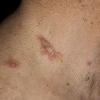
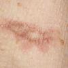
Stellate pseudoscars (see eFig. 108-4.3 in on-line edition)
|
Absence of epidermal pigmentation, altered fragmented dermal collagen
|
|
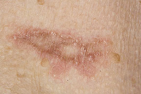
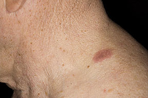
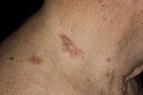
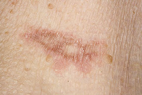
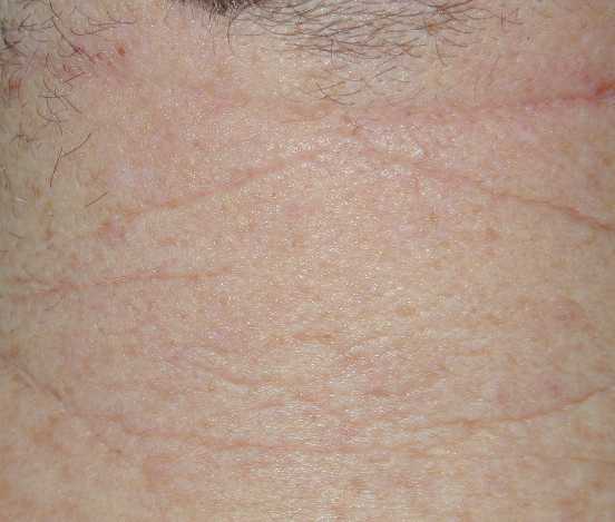
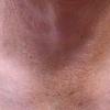
Elastosis (fine nodularity and/or coarseness)
|
Nodular aggregations of fibrous to amorphous material in the papillary dermis
|
|
Inelasticity
|
Elastotic dermis
|
|
Telangiectasia
|
Ectatic vessels often with atrophic walls
|
|
Venous lakes
|
Ectatic vessels often with atrophic walls
|
|
Purpura (easy bruising)
|
Extravasated erythrocytes and increased perivascular inflammation
|
|
Comedones
|
Ectasia of the pilosebaceous follicular orifice
|
|
Sebaceous hyperplasia
|
Concentric hyperplasia of sebaceous glands
|
|
aBasal cell carcinoma and squamous cell carcinoma also occur in photoaged skin but, unlike the table entries, affect only a minority of individuals.
|
|
|