Chromoblastomycosis
(Chromomycosis)
Chromoblastomycosis is a chronic fungal infection of the skin and subcutaneous tissues caused by pigmented or dematiaceous fungi that are implanted into the dermis from the environment. In the ensuing inflammation, they form thick-walled single cells or cell clusters (sclerotic or muriform bodies), and these may elicit a marked form of pseudoepitheliomatous hyperplasia often accompanied by trans-epidermal elimination of organisms. The infection can be caused by a number of different pigmented fungi, the most common being Phialophora verrucosa, Fonsecaea pedrosoi, F. compactum, Wangiella dermatitidis, and Cladophialophora carrionii.
The fungi that cause chromoblastomycosis can be isolated in the environment from wood, plant debris, or soil.17 The vast majority of infections are caused by F. pedrosoi and C. carrionii. As with other subcutaneous mycoses, infection follows implantation through a tissue injury. The infection is found as a sporadic condition in Central and South America, although rarely in North America. It occurs in the Caribbean region, Africa (particularly Madagascar), Australia, and Japan . It also may occur as an imported infection outside the usual endemic areas. The disease is most frequent in male rural workers
|
Macroscopic and Histopathologic Features of Mycetoma Grains
|
|
ORGANISMS
|
HEMATOXYLIN AND EOSIN SECTION APPEARANCES
|
|
Eumycetoma
|
|
|
|
Dark grains
|
|
|
|
|
Madurella mycetomatis
|
Cement present, vesicles sometimes prominent
|
|
|
|
M. grisea
|
Cement absent, compact outer layer
|
|
|
|
Leptosphaeria senegalensis
|
Cement in outer zone, dark periphery with vesicular center
|
|
|
|
Exophilia jeanselmei
|
Cement absent, often hollow
|
|
|
|
Pyrenochaeta romeroi
|
Cement lacking, compact outer layer
|
|
|
Pale grains
|
|
|
|
|
Fusarium sp., Acremonium, Scedosporium apiospermum, Aspergillus nidulans, Neotestudina rosati
|
Compact, pigment lacking, interwoven fungal filaments (S. apiospermum may have prominent vesicles)
|
|
Actinomycetoma
|
|
|
|
Pale (white to yellow) grains
|
|
|
|
|
Actinomadura madura
|
Basophilic-stained fringe in layers
|
|
|
|
Nocardia brasiliensis
|
Small, pale blue, eosinophilic fringe
|
|
|
Yellow to brown grains
|
|
|
|
|
Streptomyces somaliensis
|
Grains fractured, basophilic or pink
|
|
|
Red to pink grains
|
|
|
|
|
A. pelletieri
|
Small, basophilic layers
|
|
CLINICAL FINDINGS
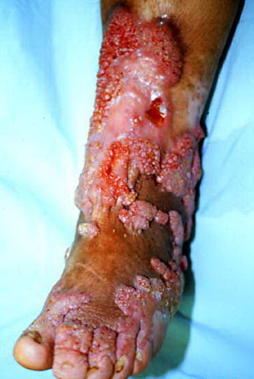
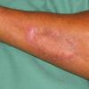
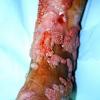
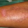
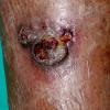
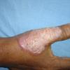
The initial site of the infection is usually on the feet, legs, arms, or upper trunk. The clinical features vary. The initial lesion is often a warty papule that expands slowly over months or years . Alternatively, lesions may be plaque-like with an atrophic center. The more common verrucous form spreads slowly and locally. Individual lesions may be very thick and often develop secondary bacterial infection. Satellite lesions around the initial site of infection are local extensions of the infection and usually are produced by scratching. Complications of chromoblastomycosis include local lymphedema, leading to elephantiasis and squamous carcinomas in some chronic lesions.
DIFFERENTIAL DIAGNOSIS
The disease must be differentiated from chronic tropical lymphedema with hyperplasia (mossy foot). Other chronic verrucous lesions, such as tuberculosis and blastomycosis, are often more extensive. The identification of organisms in the lesions of chromoblastomycosis is essential.
LABORATORY TESTS
The typical sclerotic or muriform fungal cells can be seen in skin scrapings taken from the surface of lesions, particularly areas where there is a small dark spot on the skin surface, using KOH mounts. The lesions also should be biopsied because the pathologic changes and presence of muriform cells are typical. The histology shows a mixed granulomatous response, with small neutrophil abscesses and often exuberant epidermal hyperplasia. The organisms, which are often seen either in giant cells or in neutrophil abscesses, appear singly or in small groups of brown pigmented cells, often with a single or double septum and thick cell wall.
In culture, these fungi are very similar in gross macroscopic appearance, producing black colonies with a downy surface. Their cultural identification depends on demonstrating the presence of different but specific types of sporulation, and either single or multiple sporulation mechanisms may be seen in each organism. Accurate differentiation between the different fungi may be difficult. At this stage, the choice of treatment does not depend critically on correct identification of the organisms, although there may be differences in the speed of response to azole drugs (see following section Treatment
TREATMENT
The main treatments for chromoblastomycosis are itraconazole, 200 mg daily, with or without flucytosine, 30 mg/kg qid (in a patient with normal renal function)19; terbinafine, 250 mg daily20; and, in extensive cases, intravenous amphotericin B (up to 1
mg/kg daily). Lesions can be spread by surgery, which should be used only as an adjunctive therapy after drug treatment. The local application of heat may be helpful in some instances. The responses of these fungi to different antifungal agents do not appear to differ significantly, although there is some evidence that C. carrionii responds more rapidly to terbinafine and itraconazole. In any event, treatment is continued until there is clinical resolution of lesions, which usually takes several months. Extensive lesions often respond poorly to treatment.