Chromhidrosis
Chromhidrosis is a rare condition characterized by the secretion of colored sweat. Two glands produce sweat: eccrine and apocrine glands. Eccrine glands secrete a clear, odorless fluid that serves to regulate body temperature. Apocrine glands secrete a thick, milky sweat that, once broken down by bacteria, is the main cause of body odor.
Chromhidrosis is apocrine in origin. Although apocrine glands are found in the genital, axillary, areolar, and facial skin, chromhidrosis is reported only on the face,1 axillae,2 and breast areola.3,4 Lipofuscin pigment is responsible for the colored sweat. This pigment is produced in the apocrine gland, and its various oxidative states account for the characteristic yellow, green, blue, or black secretions observed in apocrine chromhidrosis.
In contrast, eccrine chromhidrosis is rare and occurs with ingestion of certain dyes or drugs, and pseudochromhidrosis occurs when clear eccrine sweat becomes colored on the surface of the skin as a result of extrinsic dyes, paints, or chromogenic bacteria.
Approximately 10% of people without chromhidrosis have colored sweat that is regarded as acceptable and within the normal range.
Pathophysiology
Lipofuscin is a yellowish brown pigment that is normally found in the cytoplasm of relatively nondividing cells (eg, neurons). In chromhidrosis, lipofuscins are found in a higher-than-normal concentration or a higher-than-normal state of oxidation in apocrine glands. However, why some glands experience these changes is unclear. This increased level of oxidation results in the green, blue, and even black sweat seen in chromhidrosis.
The yellow, green, and blue apocrine secretions produce a yellow fluorescence under a Wood lamp (UV 360 nm), whereas the dark brown and black apocrine secretions seldom autofluoresce. Substance P is also postulated to be an important neurotransmitter in this process.
Pseudochromhidrosis is of an extrinsic etiology in which a chemical on the surface of the skin reacts with eccrine secretions and produces the color transformation.
History
History taking should include a detailed investigation of the patient's environment and lifestyle to exclude exogenous causes.
Quantities of apocrine sweat are less than those of eccrine sweat.
- Usually, patients report axillary staining of their undershirt, staining of their bra,5 or, less frequently, staining of the face or areola. Yellow is the most common color of axillary staining.
- An aura of warmth or a prickly sensation prompted by emotional or physical stimuli may precede the onset of colored sweat.
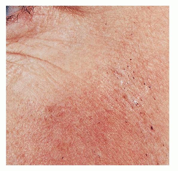
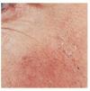
- Facial apocrine chromhidrosis is rarely described. It occurs most frequently on the cheeks and malar eminences. The secretion can often be expressed mechanically.
Physical
On careful inspection, the following signs can often be observed in chromhidrosis:
- An odorless yellow, green, blue, brown, or black and turbid secretion that can be manually expressed from apocrine-bearing skin
- Staining that is accentuated in the follicular orifices and pores
- Glistening, adherent, deeply colored flecks that appear as the secretions dry
Causes
The increased numbers of lipofuscin pigments in the secretory apocrine cells are presumed to be the cause of apocrine chromhidrosis.
Several extrinsic causes of eccrine chromhidrosis and pseudochromhidrosis include chromogenic bacteria, especially Corynebacterium species, fungi, dyes, drugs, and chemical contactants
Laboratory Studies
No significant laboratory abnormalities have been noted with apocrine chromhidrosis. The following test may help to rule out other causes:
- Determination of complete blood cell counts to exclude bleeding diathesis
- Tests of urinary homogentisic acid levels to exclude alkaptonuria
- Fungal and bacteriologic cultures to exclude infectious causes of pseudochromhidrosis
Other Tests
Wood lamp examination of colored sweat may be positive. If no sweat is produced at the time of the test, manual expression or pharmacologic stimulation with intradermal epinephrine or oxytocin (Pitocin) can be used to stimulate sweat secretion.
Clothing fibers in contact with the secretions may also fluoresce yellow-green with standard UV microscopy.13
Histologic Findings
The apocrine glands appear normal in size and morphology, but the number of glands varies. The increased number of yellow-brown lipofuscin granules is observed in the cytoplasm of secretory cells on routine hematoxylin-eosin staining. The granules are positive on periodic acid-Schiff stains and demonstrate autofluorescence under a UV excitation wavelength of 360-395 nm. Schmorl stains may also be weakly positive.
Medical Care
Apocrine chromhidrosis has no fully satisfactory cure or treatment. Patients can manually or pharmacologically empty the glands to achieve a symptom-free period of about 48-72 hours or until the glands replenish the pigment.
BOTOX® injections have been attempted in 3 cases of chromhidrosis, with mixed results. BOTOX® is predominantly used to decrease eccrine sweat in persons with hyperhidrosis. However, recent reports demonstrated improvement of facial chromhidrosis with BOTOX® lasting 19 weeks post treatment. The mechanism by which BOTOX® suppresses apocrine chromhidrosis is unclear. BOTOX® may suppress apocrine secretion by blocking cholinergic stimulation and substance P release.14,15
A few reports have described successful treatment of chromhidrosis with capsaicin cream.16,17 Capsaicin, a crystalline alkaloid found in red peppers, is commonly used for the temporary relief of pain from rheumatoid arthritis, osteoarthritis, and neuralgias. Capsaicin depletes neurons of substance P, a neurotransmitter important in apocrine sweat production. Clinical relapse occurs when therapy is stopped.
Medication
The goals of pharmacotherapy for chromhidrosis are to reduce morbidity and to prevent complications.
Counterirritants
Counterirritants may be used to treat chromhidrosis.
Capsaicin (Dolorac, Zostrix)
Derived from plants of Solanaceae family. May render skin and joints insensitive to pain by depleting substance P in peripheral sensory neurons. Use 0.025% cream.
Adult
Apply to affected area bid; not to exceed 4 applications/d
Pediatric
Administer as in adults
Documented hypersensitivity; broken or irritated skin
Pregnancy
C - Fetal risk revealed in studies in animals but not established or not studied in humans; may use if benefits outweigh risk to fetus
Precautions
Causes significant irritation and a burning sensation during first few days of use; for external use only; avoid contact with eyes; do not use tight bandage; discontinue use if condition worsens or symptoms persist for 14-28 d.
Neuromuscular blocking agents
These agents inhibit the transmission of nerve impulses at the neuromuscular junction of skeletal muscle and/or autonomic ganglia.
OnabotulinumtoxinA (BOTOX®)
Prevents calcium-dependent release of acetylcholine and produces a state of denervation at the neuromuscular junction and postganglionic sympathetic cholinergic nerves in the sweat glands.
Adult
Facial chromhidrosis: 3-5 U spaced approximately 1 cm apart over affected area; total of 10-15 U into each side of face
Pediatric
Not established