
Arteriovenous
malformation
Arteriovenous malformations (AVMs) are defects of the circulatory system that are generally believed to arise during embryonic or fetal development or soon after birth. They are comprised of snarled tangles of arteries and veins. Arteries carry oxygen-rich blood away from the heart to the body's cells; veins return oxygen-depleted blood to the lungs and heart. The presence of an AVM disrupts this vital cyclical process. Although AVMs can develop in many different sites, those located in the brain or spinal cord-the two parts of the central nervous system-can have especially widespread effects on the body.
AVMs of the brain or spinal cord (neurological AVMs) are believed to affect approximately 300,000 Americans. They occur in males and females of all racial or ethnic backgrounds at roughly equal rates.
What are the symptoms?
Most people with neurological AVMs experience few, if any, significant symptoms, and the malformations tend to be discovered only incidentally, usually either at autopsy or during treatment for an unrelated disorder. But for about 12 percent of the affected population (about 36,000 of the estimated 300,000 Americans with AVMs), these abnormalities cause symptoms that vary greatly in severity. For a small fraction of the individuals within this group, such symptoms are severe enough to become debilitating or even life-threatening. Each year about 1 percent of those with AVMs will die as a direct result of the AVM.
Seizures and headaches are the most generalized symptoms of AVMs, but no particular type of seizure or headache pattern has been identified. Seizures can be partial or total, involving a loss of control over movement, convulsions, or a change in a person's level of consciousness. Headaches can vary greatly in frequency, duration, and intensity, sometimes becoming as severe as migraines. Sometimes a headache consistently affecting one side of the head may be closely linked to the site of an AVM. More frequently, however, the location of the pain is not specific to the lesion and may encompass most of the head.
AVMs also can cause a wide range of more specific neurological symptoms that vary from person to person, depending primarily upon the location of the AVM. Such symptoms may include muscle weakness or paralysis in one part of the body; a loss of coordination (ataxia) that can lead to such problems as gait disturbances; apraxia, or difficulties carrying out tasks that require planning; dizziness; visual disturbances such as a loss of part of the visual field; an inability to control eye movement; papilledema (swelling of a part of the optic nerve known as the optic disk); various problems using or understanding language (aphasia); abnormal sensations such as numbness, tingling, or spontaneous pain (paresthesia or dysesthesia); memory deficits; and mental confusion, hallucinations, or dementia. Researchers have recently uncovered evidence that AVMs may also cause subtle learning or behavioral disorders in some people during their childhood or adolescence, long before more obvious symptoms become evident.
One of the more distinctive signs indicating the presence of an AVM is an auditory phenomenon called a bruit, coined from the French word meaning noise. (A sign is a physical effect observable by a physician, but not by a patient.) Doctors use this term to describe the rhythmic, whooshing sound caused by excessively rapid blood flow through the arteries and veins of an AVM. The sound is similar to that made by a torrent of water rushing through a narrow pipe. A bruit can sometimes become a symptom-that is, an effect experienced by a patient-when it is especially severe. When audible to patients, the bruit may compromise hearing, disturb sleep, or cause significant psychological distress.
Symptoms caused by AVMs can appear at any age, but because these abnormalities tend to result from a slow buildup of neurological damage over time they are most often noticed when people are in their twenties, thirties, or forties. If AVMs do not become symptomatic by the time people reach their late forties or early fifties, they tend to remain stable and rarely produce symptoms. In women, pregnancy sometimes causes a sudden onset or worsening of symptoms, due to accompanying cardiovascular changes, especially increases in blood volume and blood pressure.
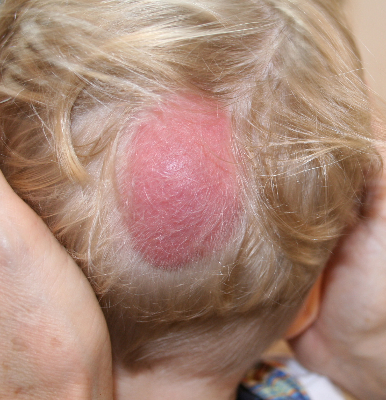
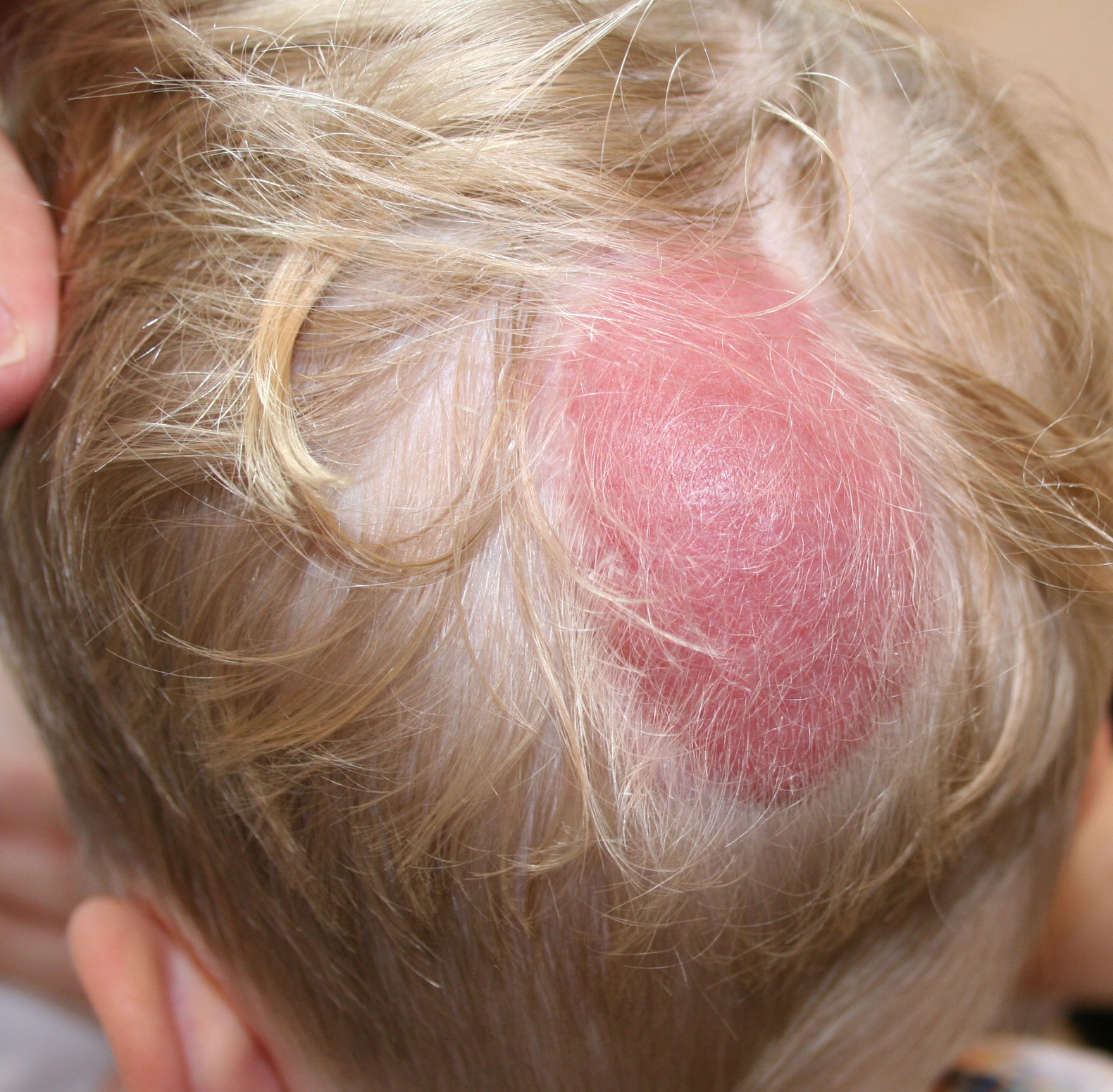
In contrast to the vast majority of neurological AVMs, one especially severe type causes symptoms to appear at, or very soon after, birth. Called a vein of Galen defect after the major blood vessel involved, this lesion is located deep inside the brain. It is frequently associated with hydrocephalus (an accumulation of fluid within certain spaces in the brain, often with visible enlargement of the head), swollen veins visible on the scalp, seizures, failure to thrive, and congestive heart failure. Children born with this condition who survive past infancy often remain developmentally impaired
How can AVMs and other vascular lesions be treated?
Medication can often alleviate general symptoms such as headache, back pain, and seizures caused by AVMs and other vascular lesions. However, the definitive treatment for AVMs is either surgery or focused irradiation therapy. Venous malformations and capillary telangiectases rarely require surgery; moreover, their structures are diffuse and usually not suitable for surgical correction and they usually do not require treatment anyway. Cavernous malformations are usually well defined enough for surgical removal, but surgery on these lesions is less common than for AVMs because they do not pose the same risk of hemorrhage.
The decision to perform surgery on any individual with an AVM requires a careful consideration of possible benefits versus risks. The natural history of an individual AVM is difficult to predict; however, left untreated, they have the potential of causing significant hemorrhage, which may result in serious neurological deficits or death. On the other hand, surgery on any part of the central nervous system carries its own risks as well; AVM surgery is associated with an estimated 8 percent risk of serious complications or death. There is no easy formula that can allow physicians and their patients to reach a decision on the best course of therapy-all therapeutic decisions must be made on a case-by-case basis.
Today, three surgical options exist for the treatment of AVMs: conventional surgery, endovascular embolization, and radiosurgery. The choice of treatment depends largely on the size and location of an AVM.
Conventional surgery involves entering the brain or spinal cord and removing the central portion of the AVM, including the fistula, while causing as little damage as possible to surrounding neurological structures. This surgery is most appropriate when an AVM is located in a superficial portion of the brain or spinal cord and is relatively small in size. AVMs located deep inside the brain generally cannot be approached through conventional surgical techniques because there is too great a possibility that functionally important brain tissue will be damaged or destroyed.
Endovascular embolization and radiosurgery are less invasive than conventional surgery and offer safer treatment options for some AVMs located deep inside the brain. In endovascular embolization the surgeon guides a catheter though the arterial network until the tip reaches the site of the AVM. The surgeon then introduces a substance that will plug the fistula, correcting the abnormal pattern of blood flow. This process is known as embolization because it causes an embolus (a blood clot) to travel through blood vessels, eventually becoming lodged in a vessel and obstructing blood flow. The materials used to create an artificial blood clot in the center of an AVM include fast-drying biologically inert glues, fibered titanium coils, and tiny balloons. Since embolization usually does not permanently obliterate the AVM, it is usually used as an adjunct to surgery or to radiosurgery to reduce the blood flow through the AVM and make the surgery safer.
Radiosurgery is an even less invasive therapeutic approach. It involves aiming a beam of highly focused radiation directly on the AVM. The high dose of radiation damages the walls of the blood vessels making up the lesion. Over the course of the next several months, the irradiated vessels gradually degenerate and eventually close, leading to the resolution of the AVM.
Embolization frequently proves incomplete or temporary, although in recent years new embolization materials have led to improved results. Radiosurgery often has incomplete results as well, particularly when an AVM is large, and it poses the additional risk of radiation damage to surrounding normal tissues. Moreover, even when successful, complete closure of an AVM takes place over the course of many months following radiosurgery. During that period, the risk of hemorrhage is still present. However, both techniques now offer the possibility of treating deeply situated AVMs that had previously been inaccessible. And in many patients, staged embolization followed by conventional surgical removal or by radiosurgery is now performed, resulting in further reductions in mortality and complication rates.
Because so many variables are involved in treating AVMs, doctors must assess the danger posed to individual patients largely on a case-by-case basis. The consequences of hemorrhage are potentially disastrous, leading many clinicians to recommend surgical intervention whenever the physical characteristics of an AVM appear to indicate a greater-than-usual likelihood of significant bleeding and resultant neurological damage.
.