|
Seborrheic keratosis, acanthotic type = التقران الدهني من النموذج الشواكي |
 |
|
Seborrheic keratoses are very common: sometimes single but often multiple. They occur mainly on the trunk and face but also on the extremities, excluding palms and soles. They usually do not appear before middle age, but are present in about 20% of the elderly (70). They are sharply demarcated, brownish in color, and slightly raised, often appearing as though stuck on the surface of the skin. Most have a verrucous surface, and a soft, friable consistency. Some have a smooth surface but characteristically show keratotic plugs. Although most lesions measure only a few millimeters in diameter, some occasionally reach a size of several centimeters. Crusting and an inflammatory base are found if the lesion has been subjected to trauma. Occasionally, small examples are pedunculated, especially on the neck and upper chest, where they resemble soft fibromas . Malignant change is rare but has been reported in association with unusual clinical appearances to the seborrhoeic keratoses .
|
|
Histopathology.
Seborrheic keratoses show a considerable variety of histologic appearances. Six types are generally recognized: irritated; adenoid or reticulated; plane; clonal; melanoacanthoma; inverted follicular keratosis; and benign squamous keratosis
|
|
Often more than one type is found in the same lesion. In addition, two clinical variants of seborrheic keratosis will be described-dermatosis papulosa nigra and stucco keratosis
|
|
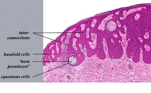
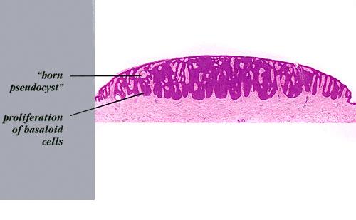
All types of seborrheic keratosis have in common hyper-keratosis, acanthosis, and papillomatosis. The acanthosis in most instances is due entirely to upward extension of the tumor. Thus the lower border of the
tumor is even and generally lies on a straight line that may be drawn from the normal
|
|
epidermis at one end of the tumor to the normal epidermis at the other end . Two types of cells are usually seen in the acanthotic epidermis: squamous cells and basaloid cells. The former have the appearance of squamous cells normally found in the epidermis; the basaloid cells are small and uniform in appearance and have a relatively large nucleus. In areas of slight intercellular edema, intercellular bridges can be easily recognized . Thus they resemble the basal cells found normally in the basal layer of the epidermis.
|
|