|
Reactive Perforating Collagenosis = الداء الكوللاجيني الثاقب الارتكاسي |
 |
 |
 |
  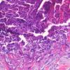  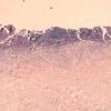 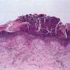 
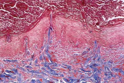
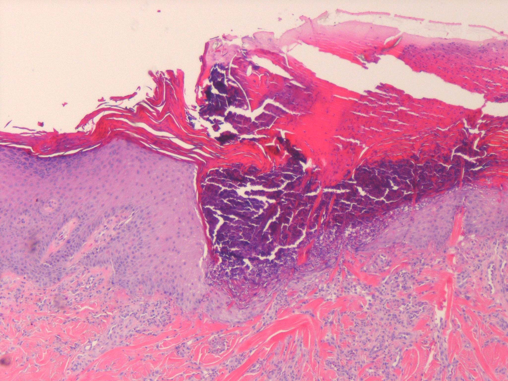
reactive perforating collagenosis
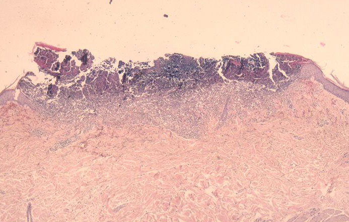
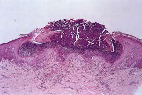
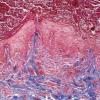
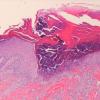
The histology varies with the stage of the reactive perforating collagenosis. Early lesions show epidermal hyperplasia associated with underlying degenerate basophilic collagen fibers. In established lesions, a cup-shaped depression of the epidermis associated with a keratin plug containing parakeratosis, inflammatory debris and collagen fibers can be seen.
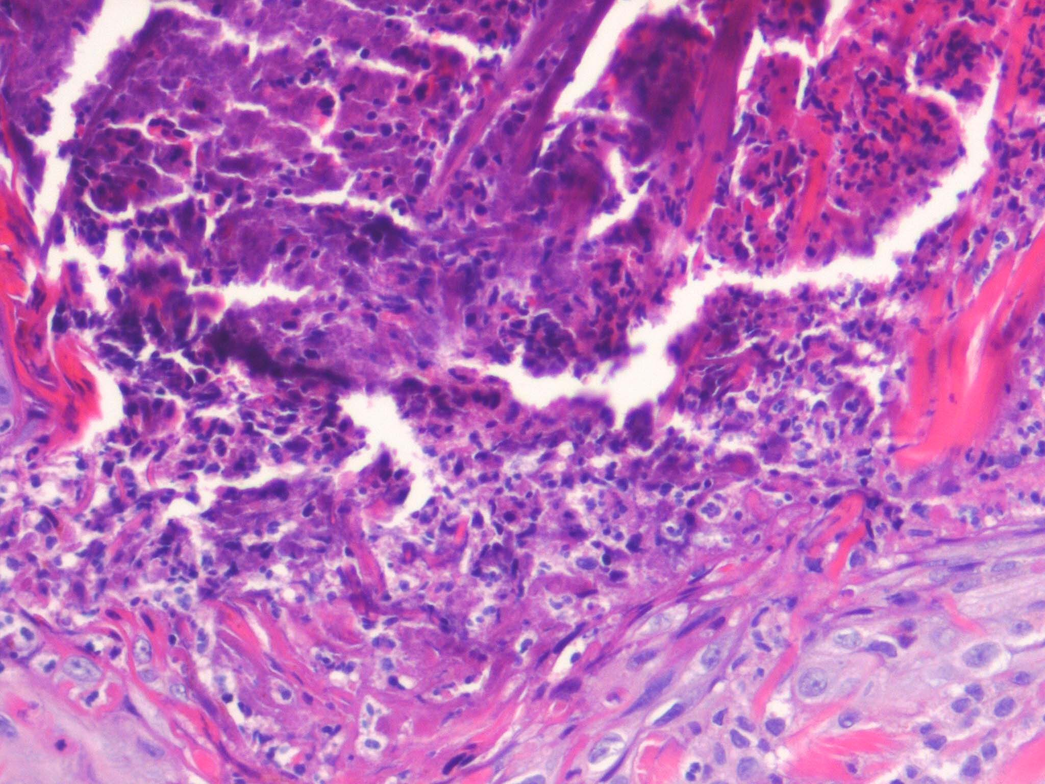
Vertically orientated basophilic collagen fibers are seen in the underlying dermis, with focal extrusion through the epidermis
The epidermis is atrophic and may show ulceration. However, at the edges of the cup-shaped invagination, the epidermis is hyperplastic. Additionally, a mild perivascular lymphohistiocytic infiltrate can be seen.
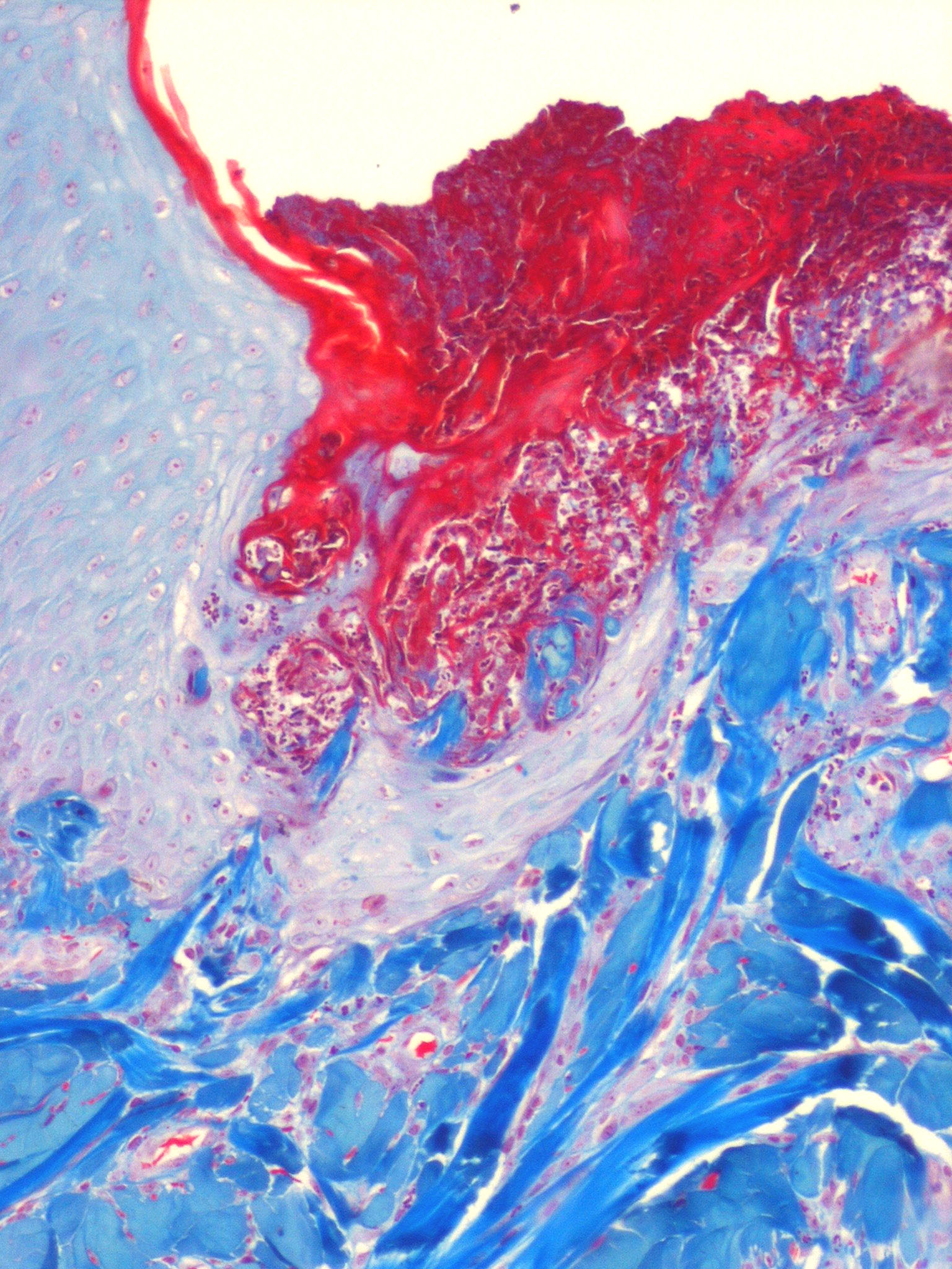
Extruded collagen fibers may be demonstrated with elastic van Gieson (EVG) staining, which stains the fibers red. No extrusion of elastic fibers should be seen (staining black with EVG).
|