| Porokeratosis = التقران الثاقب |
|
|
Porokeratosis
Porokeratosis is a morphologically distinct disorder of keratinization, characterized clinically by hyperkeratotic papules or plaques surrounded by a thread-like elevated border that expands centrifugally. Histologically, a thin column of parakeratotic cells extends throughout the stratum corneum and is seen in all variants. This distinctive histopathologic feature, known as the cornoid lamella, corresponds to the raised hyperkeratotic border evident clinically. Several clinical variants of porokeratosis have been described, some with overlapping features . Although various forms are recognized, the clinical distinction between the different variants may not be justified, because overlap occurs among the varieties. Reports of one type of porokeratosis co-existing with other forms and different types developing in multiple members of an affected family suggest more similarities than disparities, particularly in the disseminated forms. Clinical Variants of Porokeratosis
ETIOLOGY AND PATHOGENESIS Porokeratosis is a genetically heterogeneous disorder with several loci identified to date; however, the pathogenetic mechanisms remain elusive. Loci at chromosome bands 12q23.2-24.1 and 15q25.1-26 (DSAP1 and DSAP2) have been reported in familial disseminated superficial actinic porokeratoses; a further locus has been identified for disseminated superficial porokeratosis (DSP) at 18p11.3. The locus at DSAP1 corresponds to a candidate gene, SART3, that encodes a tumor rejection antigen and is thought to be involved in the regulation of messenger RNA splicing. Mutations in SART3 may thus result in altered proliferation and transformation of epithelial cells. Fine mapping of the locus at DSAP1 has also revealed mutations in another potential candidate gene, SSH1, which encodes a phosphatase that plays a key role in actin dynamics. Porokeratosis punctata palmaris et plantaris maps to a 6.9-centimorgan region at chromosome band 12q24.1-24.2 that overlaps with the region identified for DSAP1, which suggests that the two forms are allelic. The centrifugal expansion of individual lesions is postulated to reflect the migration of a mutant clone of keratinocytes. Supporting this mutant clone theory are findings of abnormal DNA ploidy and chromosomal abnormalities in lesional keratinocytes. The tumor suppressor proteins p53 and pRb are overexpressed in keratinocytes immediately beneath and adjacent to the cornoid lamella, although to date p53 mutations have not been identified. Cytogenetic abnormalities in fibroblasts, particularly on chromosome 3, have also been documented. Decreased mdm2, abnormal expression of psi-3, cytokeratins, filaggrin, and involucrin have also been reported. The increased prevalence of porokeratosis in immunosuppressed patients suggests that impaired immunity may be permissive in genetically predisposed individuals. Other reported triggering factors such as exposure to ultraviolet light, together with the increased potential for malignant transformation, highlight the
Porokeratosis of Mibelli Classic porokeratosis of Mibelli begins during infancy or childhood as asymptomatic small brown to skin-colored annular papules with a characteristic annular border (Fig. 50-1). The well-demarcated hyperkeratotic border is usually more than 1 mm in height, with a characteristic longitudinal furrow. The center of the lesion may be hyperpigmented, hypopigmented, depressed, atrophic, or anhidrotic. Lesions range in diameter from millimeters to several centimeters, but giant lesions measuring up to 20 cm may occur. Such giant porokeratoses are rare and occur predominantly on the lower leg and foot. Large lesions are associated with a higher malignant potential.33 Multiple lesions may arise; however, they are usually regionally localized and unilateral. The condition may be familial and inherited as an autosomal dominant trait. Lesions persist indefinitely.
Disseminated Superficial Actinic
Porokeratosis Disseminated superficial actinic porokeratosis (DSAP) is the most common of the porokeratoses. Lesions are characteristically uniformly small, annular, asymptomatic or mildly pruritic papules ranging from 2 to 5 mm in diameter, distributed symmetrically on the extremities. Lesions are more generalized than other forms of porokeratosis, with typically in excess of 50 lesions located predominantly in sun-exposed sites . A history of increased prominence with sun exposure is usually reported. Although widespread, lesions typically spare palms, soles, and mucous membranes. Compared with porokeratosis of Mibelli, the hyperkeratotic border is characteristically more subtle. As the lesions progress, the older, central area becomes atrophic and anhidrotic. DSAP tends to be inherited as an autosomal dominant disorder, with the earliest reported age of onset at 7 years, and is usually fully penetrant by the third or fourth decade of life. Initial reports of induction of lesions by exposure to ultraviolet light and hypersensitivity of DSAP-derived fibroblasts to x-rays have not been consistently reproduced, and the pathogenesis of DSAP remains unknown. Disseminated Superficial Porokeratosis DSP also shows an autosomal dominant pattern of inheritance and has its onset in the third or fourth decade of life. Lesions primarily are morphologically identical to those of DSAP, occur on the extremities, and are typically distributed symmetrically, but do not spare sun-protected areas as in DSAP. As with DSAP, in excess of 100 lesions may be disseminated, with a predilection for the extensor surfaces of the extremities. Notably, involvement of the face is rare in both DSAP and DSP. In both disseminated forms, there is a reported female predominance, with a female-male ratio of 3:1.
Disseminated Superficial Porokeratoses
of Immunosuppression Disseminated superficial porokeratoses of immunosuppression are recognized after renal, hepatic, and cardiac transplantation, electron beam irradiation,37 immunosuppressive chemotherapy, and the use of systemic corticosteroids; with hematopoietic malignancies38; and in the setting of human immunodeficiency virus infection.39 Porokeratosis has also been reported after bone marrow transplantation in the absence of ongoing immunosuppressive therapy, which suggests a more complex association than immunosuppression alone.40 The distribution and morphology of DSP of immunosuppression are similar to those of DASP, but a history of sun exposure is less evident. Linear Porokeratosis Linear porokeratosis is an uncommon variant that usually presents in early childhood, although congenital presentations
Disseminata
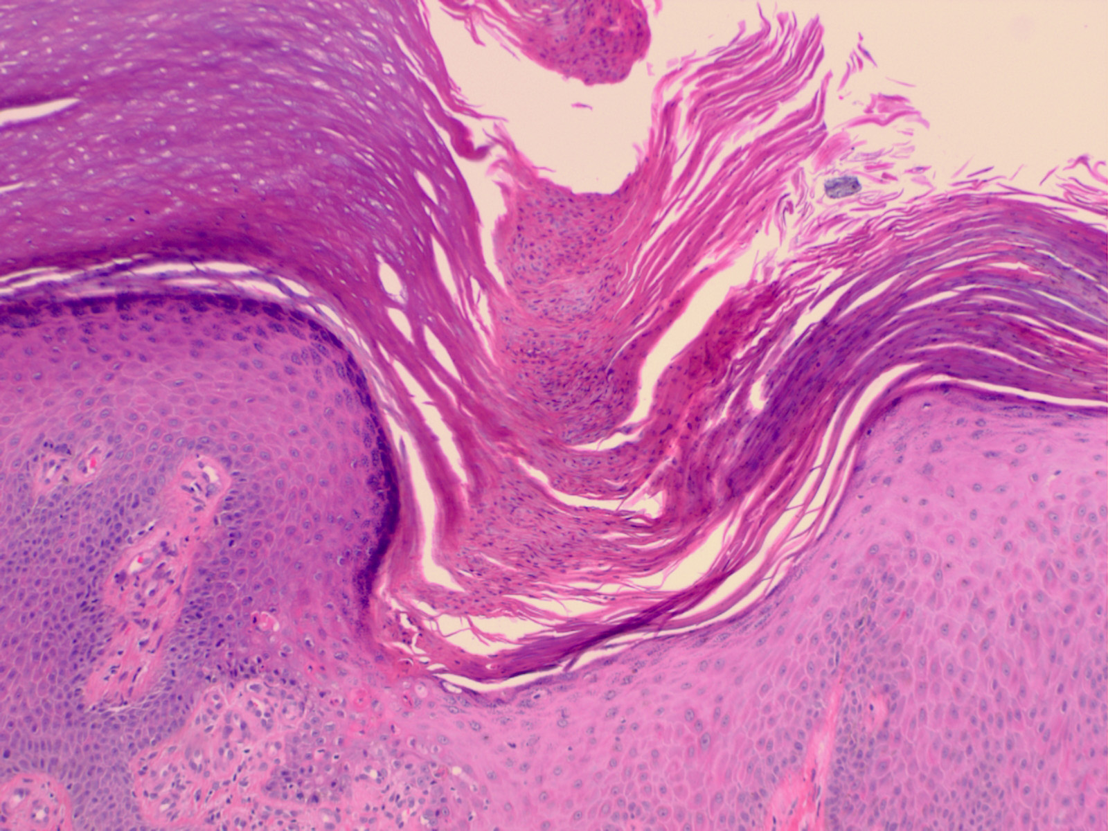
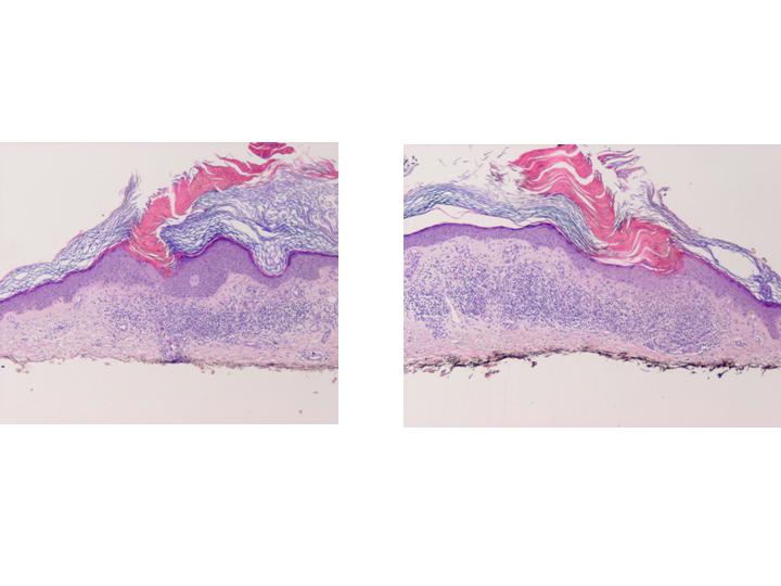
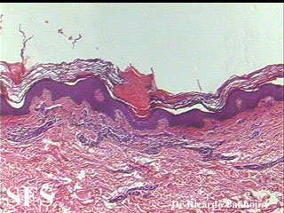
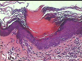
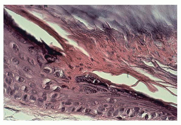
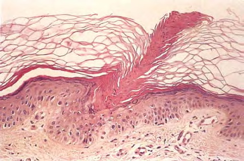
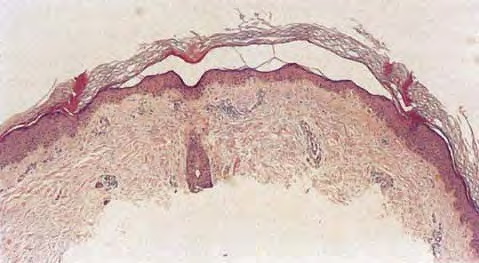
Porokeratosis palmaris et plantaris disseminata (porokeratosis punctata palmaris et plantaris) is a genodermatosis with an autosomal dominant inheritance pattern characterized by small, relatively uniform lesions that initially appear on the palms and soles. Subsequently, lesions spread to involve other parts of the body, including the mucous membranes and non-sun-exposed sites. The palmar and plantar lesions are generally more hyperkeratotic, and the characteristic longitudinal furrow along this ridge may be quite pronounced . Typically the lesions appear in during adolescence or early adulthood and are bilateral and distributed symmetrically. Porokeratosis palmaris et plantaris disseminata affects males twice as often as females. Punctate Porokeratosis Punctate porokeratosis usually appears during adolescence or adulthood and may be seen concomitantly with other types of porokeratosis. Multiple minute and discrete punctate, hyperkeratotic lesions surrounded by a thin, raised margin are present on the palms and soles. Lesions may occur in a linear arrangement, or they may aggregate to form plaques. Punctate porokeratosis must be differentiated clinically and histologically from punctate keratoderma, also referred to as punctate porokeratotic keratoderma or as porokeratosis punctata palmaris et plantaris . CDAGS Syndrome CDAGS syndrome (craniosynostosis and clavicular hypoplasia, delayed closure of the fontanel, anal anomalies, genitourinary malformations, skin eruption), also referred to as CAP syndrome (craniosynostosis, anal anomalies, and porokeratosis), is a rare genodermatosis reported in four ethnically diverse families to date. The main phenotypic features consist of craniosynostosis and clavicular hypoplasia, anal anomalies, and porokeratosis. It appears to segregate as an autosomal recessive trait, with possible linkage to chromosome band 22q12-13. The cutaneous manifestations are strikingly consistent, with the development of small, widespread porokeratotic papules from 1 month of age in affected individuals, predominantly affecting the face and extremities, with reported photoaggravation of lesions. It is postulated that dysregulation of multiple signaling pathways during embryogenesis is causative; however, no unifying pathogenetic mechanism has been identified. HISTOPATHOLOGY Histopathologic patterns are similar in all forms of porokeratosis, with the characteristic changes evident at the raised and advancing edge of the lesion. The stratum corneum is hyperkeratotic, with a thin column of poorly staining parakeratotic cells, the cornoid lamella, running through the surrounding normal-staining cells . The underlying keratinocytes are edematous with spongiosis and shrunken nuclei, and a striking dermal lymphocytic pattern may be evident. Underlying the cornoid lamella, the granular layer is either absent or markedly reduced but is of normal
DIFFERENTIAL DIAGNOSIS The classic lesions of porokeratosis are clinically distinctive, and the diagnosis is usually clinically apparent. Atypical lesions, however, may require differentiation . A cornoid lamella may be found in actinic keratoses but in actinic keratosis is distinguished by the presence of cytologic atypia. Verruca vulgaris often shows mounds of parakeratosis that are sometimes identical to cornoid lamellae, but koilocytosis is usually present. Linear porokeratosis may be clinically confused with other linear lesions, such as linear inflammatory verrucous epidermal nevus, incontinentia pigmenti (stage II), and lichen planus, none of which has a cornoid lamella. Differential Diagnosis of Porokeratosis LOCALIZED LESIONS Most Likely
Consider
LINEAR Consider
▪ TREATMENT Lesions of porokeratosis are chronic, slowly progressive, and relatively asymptomatic, although intense pruritus has been reported. Intervention is usually unnecessary, and disease surveillance is standard. If the lesions are problematic or cosmetically unacceptable, however, treatment with potent topical steroids, keratolytics, topical retinoids, topical 5-fluorouracil, imiquimod 5 percent, calcipotriol, anthralin, cryotherapy, carbon dioxide laser, pulsed dye laser, or neodymium:yttrium-aluminum-garnet laser may be considered . Treatment outcomes are variable and treatments are poorly standardized. Ablative techniques, such as curettage, excision, and dermabrasion, have also been used with variable degrees of success. Oral retinoids have been shown to give the most reproducible results, with reduction in the size and thickness of extensive lesions, although the disease typically recurs after their discontinuation. Given the association with malignancy, closer disease surveillance and a lower threshold for biopsy of suspicious lesions may be warranted in cases of giant porokeratoses and linear lesions, and in immunosuppressed individuals. Treatment for Porokeratosis
COURSE AND PROGNOSIS The porokeratoses are generally chronic and progressive, with lesions increasing in size and number with time. Typically, this process occurs over decades in porokeratosis of Mibelli but may be rapid in DSAP, particularly after sun exposure. In cases of immunocompromise, fluctuations in severity may parallel the state of immunocompetence, and there are reports of remission after removal of primary malignancy. The disease is generally considered a benign process; however, malignant degeneration may occur. Malignancy is thought to arise in 7 percent to 11 percent of individuals, although these figures are likely overestimated. Squamous cell carcinoma is the most frequently associated tumor and may be invasive. Bowen disease and basal cell carcinoma have also been reported. Spontaneous resolution of lesions has been reported, although it is exceptionally rare.
Porokeratosis Palmaris et Plantaris |
|||||||||||||||||||||||||||||||||||||||||||||||||||||||||||||