| Colloid milium =الدخنية الغروانية |
|
|
Colloid Milium
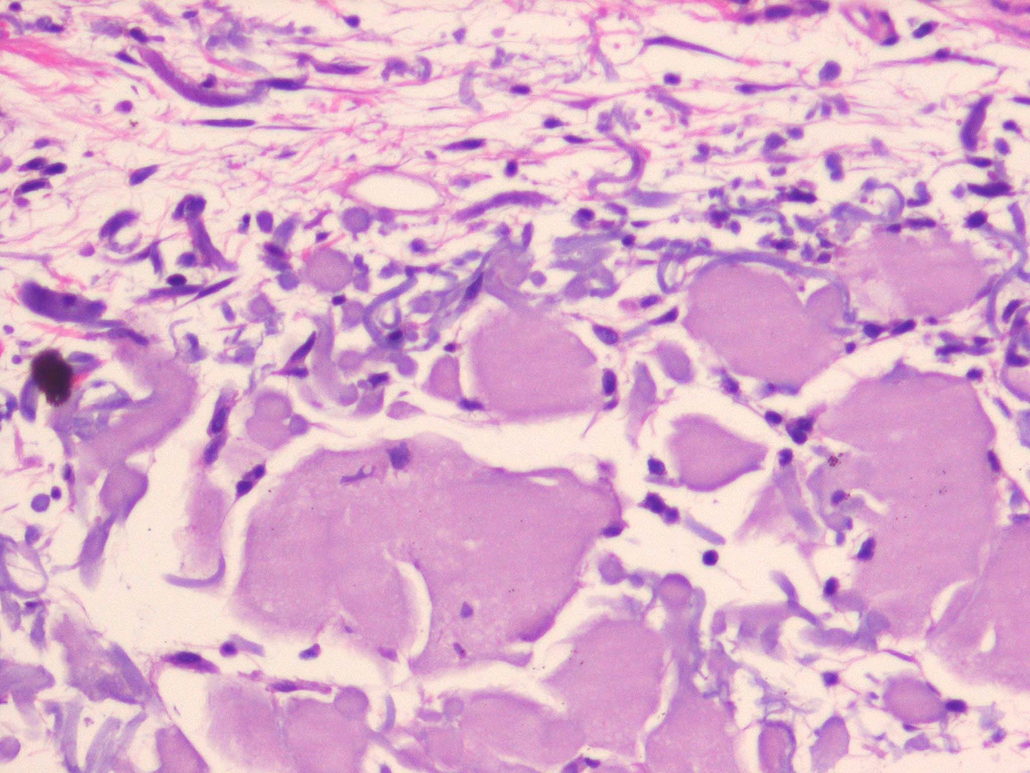
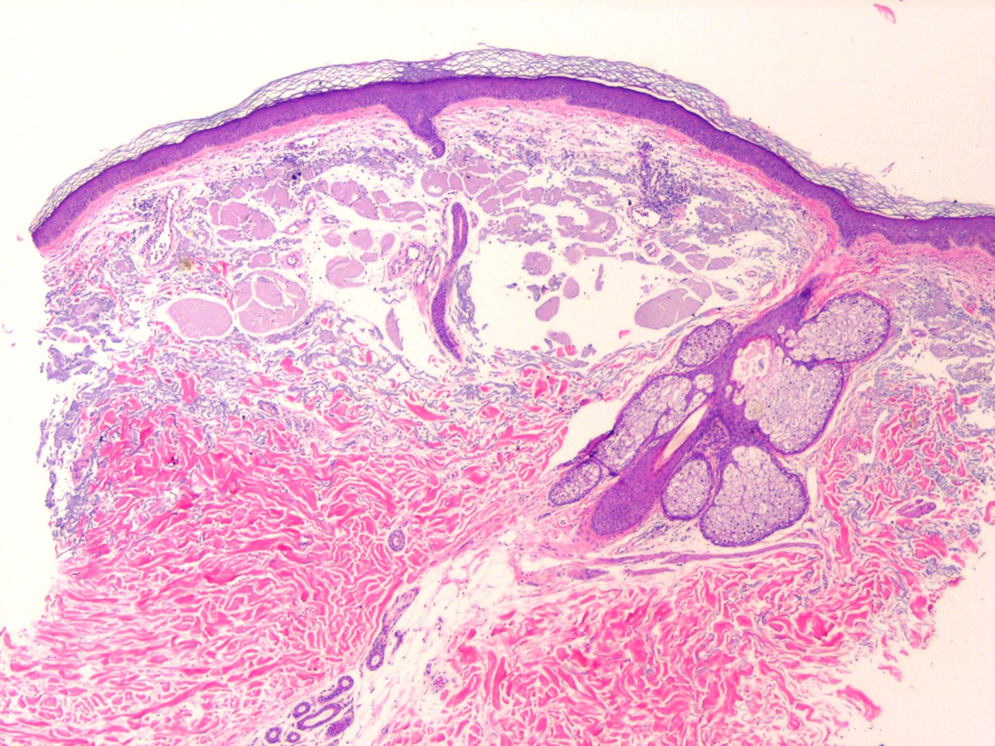
Colloid milium is a rare condition characterized by (1) the presence of multiple, dome-shaped, amber- or flesh-colored papules developing on light-exposed skin and (2) the observance of dermal colloid under light microscopy. The 4 variants are (1) an adult-onset type, (2) a nodular form (nodular colloid degeneration),1 (3) a juvenile form,2,3 and (4) a pigmented form, thought to be due to excess hydroquinone use for skin bleaching.4 Colloid milium is a degenerative condition linked to excessive sun exposure and possibly exposure to petroleum products and hydroquinone. The origin of the colloid deposition in the dermis is not certain, but it is thought to be due to degeneration of elastic fibers5,6 in the adult form and due to degeneration of UV-transformed keratinocytes in the juvenile form. Juvenile colloid milium is inherited. Colloid milium is rare, but more than 100 case reports are present in the world literature. No known figures exist on prevalence. Most cases of colloid milium persist with no natural resolution. Lesions reach their peak within 3 years, after which very few new papules occur. Colloid milium is more common in fair-skinned individuals. The adult form of colloid milium is more common in males. The rare juvenile form of colloid milium occurs before puberty. Adult colloid milium is more common in elderly patients. Papules develop gradually over the facial area and light-exposed sites. Patients with colloid milium are usually asymptomatic, but they may have transient itching in affected areas. The physical findings in colloid milium are usually limited to the skin.
Treatment For colloid milium, no treatment is available that is entirely satisfactory. Dermabrasion, cryotherapy, and diathermy have been tried with limited success. Advice about sunscreen use may also be helpful. Systemic ascorbic acid and exfoliating agents have also been tried with variable results. The Er:YAG laser may be more successful for colloid milium than dermabrasion |