|
Balloon cell nevus = وحمة الخلايا البالونية |
 |
 |
 |
|
Balloon cell nevi are histologic curiosities that possess no clinical features by which they can be differentiated fromothernevi. They are quite rare.
|
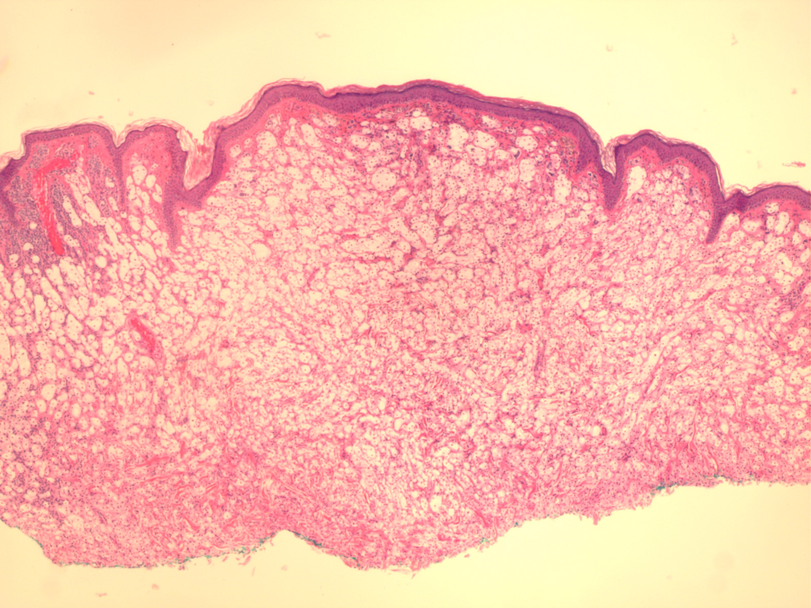
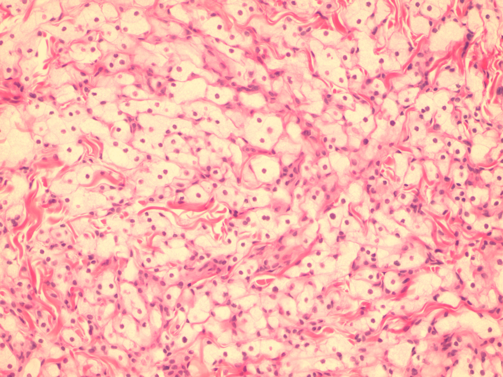
Histopathology.
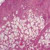
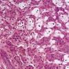
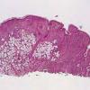
Balloon cells may be seen within the epidermis singly or in groups or may be absent from the epidermis. In the dermis, they lie arranged in lobules of varying size, often with an admixture of ordinary nevus cells and often with transitional forms between the ordinary and ballooned nevus cells . The balloon cells may be multinucleated and are considerably larger than ordinary nevus cells. Their nuclei are small, round, and usually centrally placed. Their cytoplasm appears empty, finely granular, or vacuolated, often with a few small melanin granules. There may be melanophages that are solidly packed with pigment. Stains for lipids, glycogen, and acid or neutral mucopolysaccharides are negative in the balloon cells. Electron microscopic examination reveals in balloon cells numerous large vacuoles formed by enlargement and coalescence of degenerating melanosomes . Balloon cell nevus is differentiated from balloon cell melanoma by the usual criteria. The large adipocytes present in some intradermal nevi as a result of fatty infiltration or stromal metaplasia differ from balloon cells by routine histology by having a flattened nucleus located at the periphery of the cell. In the differentiation from clear cell hidradenoma and other clear cell tumors, the absence of periodic acid-Schiff-positive glycogen and keratin in balloon cell nevus might be helpful; balloon nevus cells also stain for S100 protein, and although eccrine neoplasms (and adipocytes) may also express this marker, they will also usually be positive for Melan-AIMART-1 and negative for keratin markers.
|