| Apocrine hidrocystoma =الكيسوم العرقي المفترز |
|
|
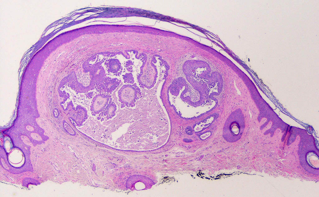
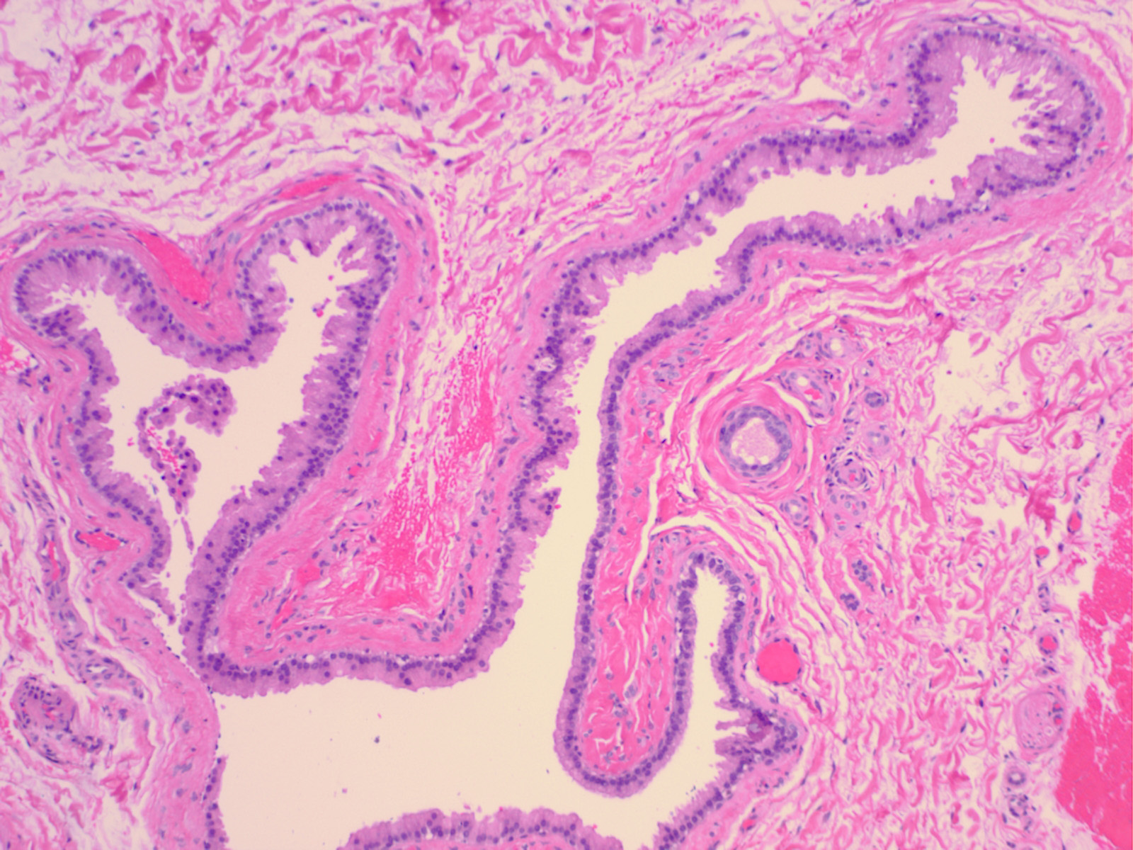
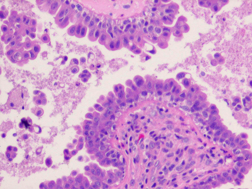
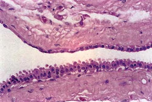
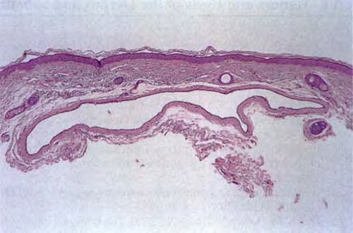
Apocrine hidrocystoma The clinical appearance of a pea-sized cyst near the inner canthus of the eye, which contains a thin clear or pigmented fluid, suggests an apocrine hidrocystoma; however, histologic examination often is required to establish a specific and definitive diagnosis. Upon histologic examination, apocrine hidrocystomas show large unilocular or multilocular cystic spaces within the dermis (see the image below). Apocrine hidrocystomas are more likely to be multilocular than the closely related eccrine hidrocystoma The cyst wall is lined by apocrine-type secretory epithelium. The innermost layer of the wall is composed of a single (occasionally double) layer of cuboidal-to columnar-shaped cells. The nuclei of these cells are positioned basally. The outer layer of cells composing the cyst wall is formed by myoepithelial cells in which the long axes run parallel to the cyst wall. |