|
Angiokeratoma = القرنوم الوعائي |
 |
 |
 |
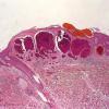  
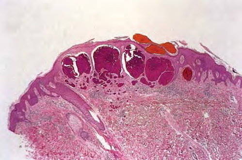
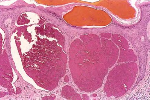
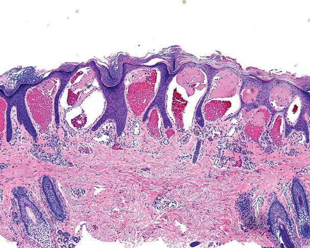
Angiokeratoma Corporis
DiffusumFabry
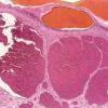
Histology shows numerous, dilated, thin-walled, endothelial-lined, blood-engorged capillaries in the papillary dermis, with an overlying hyperkeratotic epidermis. Careful inspection may reveal cytoplasmic vacuoles containing lipid in the endothelial cells, fibroblasts, and pericytes. However, in most patients, histologic findings essentially are identical to those of other angiokeratomas.
Endomyocardial biopsy findings of the heart can demonstrate sarcoplasmic vacuolization of cardiac muscle cells under light microscopy and lamellated zebra bodies in the cytoplasm under electron microscopy.
Electromicroscopy investigation can show stromal cells in hemizygous tissue and endothelial and smooth muscle cells in heterozygous tissue; cells contain membrane-bound inclusions with a lamellar structure (ie, inclusion bodies with a zebralike appearance).
|