|
Acne keloidalis nuchae =العد الجدري |
 |
 |
 |
 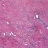 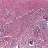 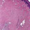
Acne keloidalis nuchae
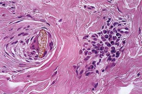
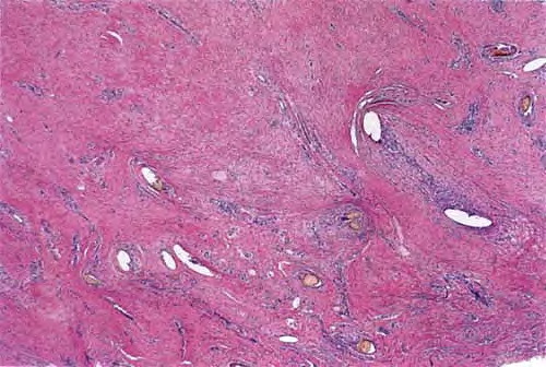
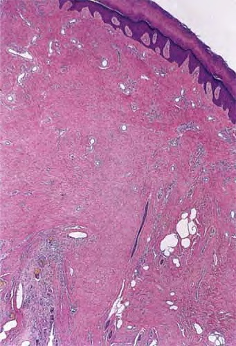
The histological findings vary depending on the timing of the biopsy. The initial infiltrate is primarily composed of neutrophils and lymphocytes that are distributed around the lower infundibulum and isthmus of the hair follicle. Subsequently, the follicle and sebaceous glands are destroyed, with liberation of the naked hair shafts into the dermis. Acute and granulomatous inflammation surrounds the free hair shafts, and, ultimately, fibrosis ensues. Scarring alopecia ensues in long-standing lesions, marked by dermal fibrosis associated with numerous plasma cells. True keloidal collagen is typically not a feature.
Often, acute and chronic inflammation may be present in the same region, because new lesions often develop adjacent to chronic lesions. Sinus tracks can be identified in long-standing lesions. Intact hair follicles at the margins may exhibit polytrichia, with more than one hair shaft noted in a single follicle, but this is physiologic for the occiput.14 Individual early papules may also demonstrate ingrown hairs, and these may be seen clinically in patients without progressive scarring alopecia.
|