|
Tattoo Reactions
Clinically apparent inflammatory reactions to permanent tattoos, although uncommon, are now seen with greater frequency due to the rise in popularity of tattoos. They have been observed most commonly with red dyes containing mercuric sulfide, such as cinnabar (Chinese red). More recently, there has been a move away from using mercury-containing dyes toward the use of dyes containing other red pigments, such as ferric hydrate (sienna or red ochre), cadmium selenide (cadmium red), and organic dyes, but such mercury-free red dyes may also produce adverse reactions . Reactions have also been reported with chrome green , cobalt blue , purple manganese salts , yellow cadmium sulfide , and iron oxide . In some instances, an allergic response to the pigment has been suggested by a positive patch test.
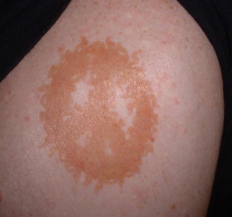
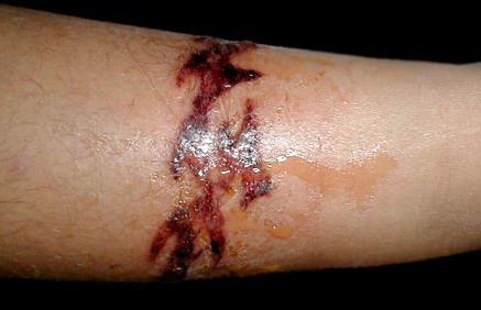
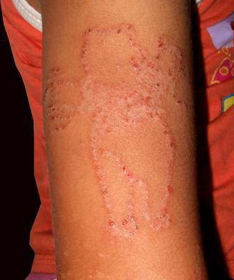
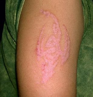
Reactions to "temporary" tattoos are rare. Usually comprised of black commercial henna, these tattoos are painted onto the skin surface. The most common reaction is allergic contact dermatitis, but a lichenoid dermatitis, a scarring reaction, and hypopigmentation have been reported .
Tattoos may also occur due to pigmented materials accidentally implanted in the skin, such as graphite, or due to solutions employed for hemostasis , particularly Monsel's solution (ferric subsulfate).
Histopathology.
Permanent tattoos that are not clinically inflamed show irregularly shaped granules of dye that are located within macrophages and extracellularly in the dermis .
Inflammatory reactions in clinically inflamed permanent tattoos mayor may not be granulomatous. Photoexacerbation has been described with reactions to red pigments and yellow pigments . Nongranulomatous reactions include a perivascular lymphocytic infiltrate with pigment-containing macrophages , a lichenoid response, which in some instances may resemble lichen planus or hypertrophic lichen planus , and a pseudolymphomatous picture with a dense, nodular or diffuse, predominantly lymphocytic infiltrate that also contains histiocytes and coarse tattoo pigment granules .
.
Granulomatous reactions may be either of the sarcoidal type or the foreign-body type . A tuberculoid pattern has also been described in response to cobalt blue, but this may have been due to a mycobacterial infection . The granulomatous responses show tattoo granules scattered throughout the infiltrate. In the sarcoidal type, the infiltrate contains nodules of epithelioid histiocytes , and in the foreign-body type, there are obvious multinucleated histiocytes of the foreign-body type. A sparse or dense lymphocytic infiltrate may be present. In the sarcoidal type of reaction, regional lymph nodes may also show tattoo granules . There are some reports of patients with sarcoidal granulomas in their tattoos who also had pulmonary disease , uveitis , or erythema nodosum , suggesting a systemic hypersensitivity response to the tattoo, or true sarcoidosis.
Traumatic graphite tattoos show black granules free in the dermis and sometimes within histiocytes . Monsel's tattoos, also typically seen in conjunction with scars, show multinucleate histiocytes containing coarse, brown, refractile pigment , which is positive on staining for iron. Larger, brown, extracellular jagged aggregates of pigment are suggestive of Monsel's tattoo . Ferruginization of collagen bundles is typical, and a proliferation of spindled fibrohistiocytic cells may also occur.
Electron microscopic examination of tattoos without an allergic reaction shows that most tattoo granules are located within macrophages, where they often lie within membrane-bound Iysosomes. In addition, some tattoo granules are found free in the dermis .
|