| Erythema dyschromicum perstans = الحمامى الدائمة بخلل التلون |
|
|
erythema dyschromicum perstans erythema dyschromicum perstans was called dermatosis ceniciento, meaning ashy dermatosis, because of its ashy bluish gray color. The term erythema dyschromicum perstans is credited to Marion B. Sulzberger, who suggested it when examining Convit's2 patients in Caracas
the narrow red border (which is often hard to find), represents the active lesions. This is why I suggested a name which contains the term "erythema" and which also suggests the variety and persistence of the final dyschromias. The descriptive term ashy dermatosis was also used as a designation for their coloration. In South America, another name, erythema chronicum figuratum melanodermicum, is also used. Erythema dyschromicum perstans (ashy dermatosis) is a distinct and somewhat controversial cutaneous eruption that may be best regarded as a form of lichen planus or lichen planus actinicus.
PathophysiologyThe etiology of erythema dyschromicum perstans is unknown, but many consider erythema dyschromicum perstans to be a variant of lichen planus actinicus. A variety of predisposing factors have been cited. These include ingestion of ammonium nitrite, an intestinal parasitosis caused by nematodes (whipworm infection, control of which produced erythema dyschromicum perstans remission), orally administered radiographic contrast media, and, possibly, an occupationally associated cobalt allergy in a plumber. One case may be particularly revealing, that of a 13-year-old rural northern European truant who repeatedly ingested small amounts of a fertilizer, ammonium nitrate, to induce erythema dyschromicum perstans and avoid school. Chlorothalonil exposure among banana farm workers is another possible cause of erythema dyschromicum perstans.6
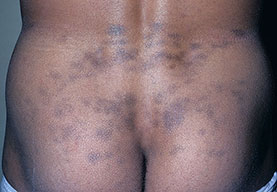
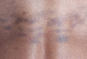
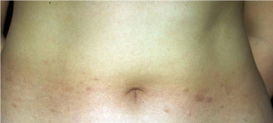
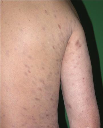
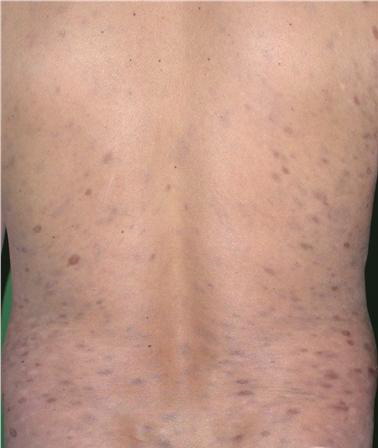
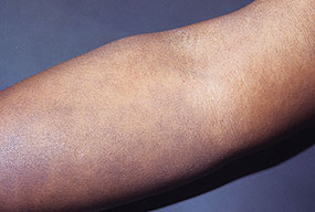
HistoryErythema dyschromicum perstans has a slow onset and is unlikely to resolve spontaneously.13 The clinical course of childhood (prepubertal) may differ from that of adults; erythema dyschromicum perstans may be more likely to resolve within 2-3 years. A proposed clinical classification has been devised, dividing ashy dermatoses from erythema dyschromicum perstans with the former lacking erythematous borders, and having a third category for simulators such as lichen planus variants, and medication-induced melanodermas.15 Erythema dyschromicum perstans is an asymptomatic eruption of oval, polycyclic, or irregularly shaped, gray-blue hyperpigmented macules on the trunk, the arms, the face, and the neck. It begins as ash-colored macules, sometimes with an erythematous or elevated border (see the image below). No systemic symptoms or associations exist. Erythema dyschromicum perstans has asymptomatic, gray-blue hyperpigmented patches of variable shape and size and an elevated erythematous border in the early stages . The eruption is symmetrically distributed on the face, the trunk, and the upper extremities. The oral cavity and the genitals are spared.
Laboratory StudiesAll cases of erythema dyschromicum perstans (EDP), to date, have resulted in negative laboratory study results, which include the following:
Imaging StudiesRadiographic studies in erythema dyschromicum perstans patients have not shown abnormalities. Histologic FindingsThe biopsy specimen is obtained as much to rule out other diagnoses as to confirm that of erythema dyschromicum perstans because the erythema dyschromicum perstans histologic pattern is relatively nonspecific. One should attempt to obtain a biopsy sample of the border of an active macule, which usually demonstrates mild basal cell layer vacuolar degeneration overlying an upper dermis with a mild perivascular mononuclear cell infiltrate and increased melanophages. Immunopathologic study of erythema dyschromicum perstans shows immune-associated (Ia) antigen expression in keratinocytes and strong OKT 4 and OKT 6 staining of Langerhans cells. It also shows dermal infiltration by T lymphocytes of both helper-inducer and suppressor-cytotoxic phenotypes, a pattern commonly seen with lichen planus. CD36 expression is evident in the viable upper epidermis on lesional keratinocytes, which may imply a delayed hypersensitivity reaction. Beneath, in the dermis, the cellular infiltrate has been found to express CD69 and the cytotoxic cell marker CD94. In addition, as with lichen planus, the colloid bodies stained immunoglobulin G positive
Medical CareMany therapeutic options are available for erythema dyschromicum perstans (EDP), but few have been effective, except for clofazimine. In one series of 8 patients, 7 had a good or excellent response to clofazimine administered either 100 mg every other day to patients weighing less than 40 kg or 100 mg every day to patients weighing more than 40 kg. This medication was continued for 3 months, then reduced to 200 mg/wk and 400 mg/wk, respectively. The one remaining patient had only a marginal response. One study found some improvement in early cases, but no cures were reported. This medication seems to have a valuable effect on the inflammatory phase of erythema dyschromicum perstans.Clofazimine is a lipophilic rhimophenazine dye with both antimicrobial and anti-inflammatory properties originally developed to treat tuberculosis. Although its mechanism of action is unclear, it seems to exert its main effect upon neutrophils and monocytes in a variety of ways, such as stimulating phagocytosis and release of lysosomal enzymes.20 Clofazimine is administered orally, with improved bioavailability when taken with food. It concentrates in lipid-rich tissues, including the reticuloendothelial system, the intestine, the breasts, and the liver. Its half-life is 70 days. Isoniazid increases its serum levels and enhances its urinary excretion. Its most common adverse effects are in the skin, the gut, and the eye. It gives a temporary orange discoloration of the skin and the eye (ie, cornea, conjunctivae); it also may produce ichthyosis. Its most serious adverse effect is crystal deposition in the gut that produces a potentially fatal enteropathy. This rare complication is associated with months of high-dose (>100 mg/d) therapy. Nausea and diarrhea are more common. Splenic infarction and eosinophilic enteritis are also rare adverse effects. Many other therapeutic modalities have been attempted, none with satisfactory results. These include ultraviolet exposure, ultraviolet avoidance, antibiotics, antihistamines, griseofulvin, chemical peels, antibiotics, corticosteroids, vitamins, isoniazid, chloroquine, and psychotherapy. A patient from Turkey was described to have responded remarkably well to treatment with dapsone.21 MedicationThe goals of pharmacotherapy are to reduce morbidity and to prevent complications. LeprostaticsHave both antimicrobial properties and anti-inflammatory properties. They were originally developed to treat tuberculosis. Clofazimine (Lamprene)Lipophilic rhimophenazine dye with antimicrobial and anti-inflammatory properties originally developed to treat tuberculosis. Inhibits mycobacterial growth, binds preferentially to mycobacterial DNA. Has antimicrobial properties, but mechanism of action unknown. Adult<40 kg: 100 mg PO qod; continue for 3 mo then reduce to 200 mg/wk PediatricNot established Dapsone may inhibit anti-inflammatory activity; avoid concurrent administration of clofazimine with aluminum-magnesium-containing antacids due to decreased absorption; concurrent use of phenytoin and clofazimine may result in reduced phenytoin efficacy; concurrent administration of small quantities of orange juice with clofazimine may result in modest reduction of clofazimine relative bioavailability Documented hypersensitivity PregnancyC - Fetal risk revealed in studies in animals but not established or not studied in humans; may use if benefits outweigh risk to fetus PrecautionsMost noticeable adverse effect is skin discoloration (red to purple-black), which fades slowly on withdrawal; secretions discolored; urine becomes red; ichthyosis of shins and forearms may be prominent Dapsone (Avlosulfon)Bactericidal and bacteriostatic against mycobacteria; mechanism of action is similar to that of sulfonamides, in which competitive antagonists of PABA prevent formation of folic acid, inhibiting bacterial growth Adult50-300 mg PO qd PediatricNot established
|