|
Epidermolysis bullosa simplex, Dowling-Meara type =انحلال البشرة الفقاعي البسيط لدولينغ ميارا |
 |
 |
 |

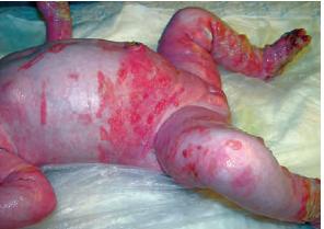
Epidermolysis bullosa simplex, Dowling-Meara type
The distribution and morphology of tonofilament (TF) clumps were examined by light and electron microscopy in skin samples from a total of 17 patients with the Dowling-Meara (DM) form of epidermolysis bullosa simplex (EBS). TF clumps extending from the basal to the upper-spinous epidermal layer were seen in all lesional skin samples and in the majority of peri-lesional and non-lesional skin samples. TF clumps were also noted in adnexal epithelia, including outer hair root sheaths, sweat ducts, and sebaceous glands. Cultured keratinocytes from two patients also demonstrated characteristic TF clumps. All these epithelial cells have in common their expression of the keratin pair K5 and K14. Post-embedding immunogold electron microscopy using antibodies to K5, K14, and K10 showed similar expressed keratins in DM-EBS skin from four patients compared with normal skin, with K5 and K14 predominantly in the basal cell layer and K10 in the suprabasal layers. The clumped TF in DM-EBS samples were labeled strongly with anti-K5 and K14 antibodies in the basal and suprabasal layers. In contrast, the suprabasal clumps were only slightly reactive with anti-K10 antibodies and labeling was usually restricted to the periphery of the clumps. We conclude that DM-EBS is associated with an intrinsic abnormality of the keratin-filament network involving the K5 and K14 pair that is likely to result in impaired resistance of basal epidermal cells to external shearing forces, leading to the characteristic intraepidermal blisters. DM-EBS may become the first genetic skin disease to be recognized as having a specific keratin abnormality
Two cases of the Dowling-Meara type of epidermolysis bullosa simplex (EBS) are described. Both had severe blistering at birth, which improved gradually with age. Vesicles and small bullae clustering in a herpetiform fashion were seen in both cases. One showed mild pincer deformity of the nails. and in the other the nail plates were shed after subungual blistering, but regrew without deformity, Histopathology and ultrastructural study showed cytolysis of the basal cells in both cases, but ultrastructurally different forms of tonofilament clumps were present in epidermal keratinocytes. In one case there was typical round clumping of tonofilaments, and in the other a whisk-type clumping of tonofilaments. Cultured keratinocytes from the former produced round clumps of keratin filaments, but those from the latter did not. Review of previous reports of Dowling-Meara EBS revealed that cases could also be divided into two groups in terms of the type of tonofilament clumping at an ultrastructural level. The possibility of subtyping of Dowling-Meara EBS, and possible mechanisms of the blistering in this disease are discussed.
|