| Eosinophilic fasciitis = التهاب الصفاق بالحمضات |
|
|
Eosinophilic fasciitis
Eosinophilic fasciitis (EF) is a rare, localized fibrosing disorder of the fascia. The etiology and pathophysiology are unclear.
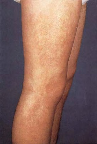
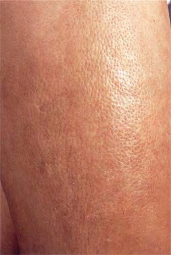
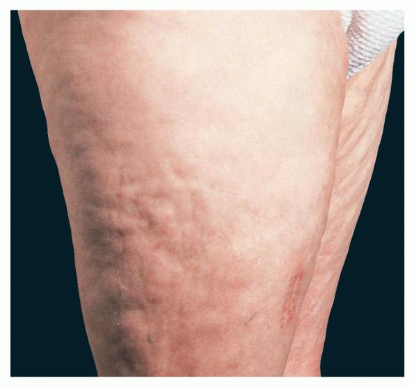
In 1974, Shulman provided an early description of eosinophilic fasciitis as a disorder characterized by peripheral eosinophilia and fasciitis that could be differentiated from scleroderma by the distinctive pattern of skin involvement that spares the digits, involves fascia rather than dermis, and is not accompanied by Raynaud phenomenon.1 Since 1974, over 250 patients with eosinophilic fasciitis have been reported.2 Despite this, the current understanding of the disease relies on a relatively few large case series and multiple case reports. Therefore, the understanding of key aspects of the disease continues to evolve. The etiology of eosinophilic fasciitis remains unknown, although many possible triggers and disease associations have been suggested. Some aspects of pathophysiology have been elucidated; however, a more complete understanding has yet to develop. The available literature has generated a broader clinical image of the condition, but fascial thickening in the setting of eosinophilia, elevated sedimentation rate, and hypergammaglobulinemia remain critical elements of the syndrome. Visceral involvement in eosinophilic fasciitis is generally absent, a finding that helps differentiate eosinophilic fasciitis from systemic sclerosis and other differential considerations. However, an association with several hematologic diseases is recognized and frequently carries a grave prognosis. The diagnosis of eosinophilic fasciitis is suspected in a patient presenting with characteristic skin changes and consistent laboratory findings. It is confirmed with full-thickness biopsy or characteristic MRI findings. Eosinophilic fasciitis is generally corticosteroid-responsive, and initial treatment regimens are based on this therapy. Multiple additional agents have been used in steroid-refractory disease. The evidence for many of these agents is anecdotal, and there is no general consensus regarding the best agent for treatment of steroid-resistant disease or cases refractory to steroid withdrawal. PathophysiologyAlthough the etiology of eosinophilic fasciitis is unknown, studies have shed light on some of the mechanisms involved in its pathogenesis. In general, the pathophysiology underlying eosinophilic fasciitis is postulated to involve an inflammatory response resulting in an activated inflammatory cell infiltrate of affected tissues and subsequent dysregulation of extracellular matrix production by lesional fibroblasts. Viallard et al (2001) demonstrated that, when stimulated, peripheral blood mononuclear cells of eosinophilic fasciitis patients produce significantly higher amounts of 5 cytokines, including interleukin (IL)–5 and interferon (IFN)–gamma.3 IL-5 is known to activate mature eosinophils and to stimulate eosinophil chemotaxis, growth, and differentiation. IFN-gamma activates tissue macrophages and T cells. The findings of Dziadzio et al (2003) support increased levels of IL-5 in eosinophilic fasciitis, in addition to increased levels of transforming growth factor (TGF)–beta, another fibrogenic cytokine.4 Toquet et al (2003) investigated the phenotype of the lesional inflammatory cell infiltrate in patients with eosinophilic fasciitis and demonstrated a predominance of macrophages, CD8+ lymphocytes, and few eosinophils.5 Pathologic specimens from patients with eosinophilic fasciitis demonstrate increased numbers of eosinophils, especially early in the disease course. Taken together, the findings of these studies suggest a mechanistic framework marked by a proinflammatory and fibrogenic cytokine response with resultant tissue inflammatory cell infiltration. In the tissues, the end effector cell of fibrosis is the fibroblast. Fibroblasts from lesional tissue of patients with eosinophilic fasciitis produce excess collagen in vitro and display elevated TGF-beta and type 1 collagen mRNA levels when examined via in situ hybridization with specific cDNA.6,7 Therefore, the pathogenesis appears to involve the concomitant increase in the expression of genes for TGF-beta and extracellular matrix proteins in fibroblasts in the affected tissues. Mori et al (2002) suggested that an autocrine stimulatory loop involving major basic protein, a product of eosinophil degranulation, IL-6, which enhances collagen production and is induced my major basic protein, and TGF-beta could account for the progressive fibrosis seen in several eosinophil prominent disorders.8 Other studies showed elevated levels of serum manganese superoxide dismutase and tissue metalloproteinase 1 (TIMP-1) in eosinophilic fasciitis, suggesting a role in pathogenesis and providing a possible marker of disease activity.9 Fasciitis may be a common manifestation of various pathophysiologic processes associated with eosinophilia. The existence of primary and secondary forms of fasciitis has recently been suggested. Understanding the mechanisms involved in the development of fascial inflammation and fibrosis in these conditions may yield insights into the pathogenesis of other fibrotic skin diseases.
History
Physical
CausesThe etiology of eosinophilic fasciitis is unknown. The clinical manifestations of eosinophilic fasciitis are the result of an inflammatory response in the affected tissues. As explained above, our current understanding of eosinophilic fasciitis relies on a relatively few case series and case reports. As such, many etiologic factors have been suggested with varying degrees of supporting evidence. It may be possible that any of these factors, alone or in combination, could initiate this inflammatory response. Several possible triggers have been reported with some consistency. A preceding history of vigorous exercise or trauma has been reported in 30%-50% of patients.15,17 Multiple drugs have also been implicated, including simvastatin, atorvastatin, and phenytoin.20,21,22 Several cases have demonstrated positive Borrelia serologies. The significance of this finding continues to be debated. Spirochetes were visualized by silver stain in 4 patients in one study.23 These findings have not been repeated. It has been suggested that positive serology for Borrelia represents an epiphenomenon among cases from Borrelia -endemic areas and is insufficient evidence of infection and therefore does not support a causal association.24 Eosinophilic fasciitis shares clinical similarities, as well as key differences, with eosinophilia-myalgia syndrome. Some studies have suggested an association between l-tryptophan ingestion and eosinophilic fasciitis.25,26 Despite this, there is no consistent association between l-tryptophan or other dietary exposure and eosinophilic fasciitis. As evidence, l-tryptophan use was significantly associated with dyspnea, an uncommon finding in eosinophilic fasciitis cases. In another instance, a patient with eosinophilic fasciitis had used l-tryptophan for several years but had started a formal exercise program 2 weeks prior to disease onset. Multiple additional etiologic triggers have been suggested by single or infrequent case reports. As with etiology, eosinophilic fasciitis has been associated with several diseases.27 Hematologic diseases have been consistently reported and are supported by large case series and case reports.15,17,28 The spectrum of associated hematologic disease is broad and includes aplastic and hemolytic anemia, thrombocytopenia, myeloproliferative disorders, myelodysplastic disorders, lymphoma, leukemia, monoclonal gammopathy of undetermined significance (MGUS), and multiple myeloma.28,29,30 An association with thyroid disease has been reported in several cases.31 Eosinophilic fasciitis has rarely been linked to solid-organ tumors and primary biliary cirrhosis, in addition to several other diseases. These disease associations may suggest a shared pathophysiology of cellular dysregulation and/or autoimmunity.
Laboratory StudiesCharacteristic laboratory findings of eosinophilic fasciitis (EF) include the following: Additional laboratory findings of eosinophilic fasciitis include the following:15,17,36 Imaging StudiesOther TestsProceduresHistologic FindingsInflammation, edema, thickening, and sclerosis of the fascia are hallmarks of eosinophilic fasciitis. Acute findings include infiltration of deep fascia and an adjacent subcutis layer with lymphocytes, plasma cells, histiocytes, and eosinophils. Distribution of the eosinophils in the fascia may be focal, and a close relationship appears to exist between blood and tissue eosinophilia. In the deeper portions of the panniculus, a similar infiltrate is found in the fibrous septa and at the periphery of the fat lobules. Deep in the fascia, the inflammatory infiltrate can extend into the epimysium, perimysium, and endomysium. In addition, vascular cuffing with lymphocytes and plasma cells is often seen.7,11,48 As the disease progresses, inflammatory changes are replaced by generalized sclerosis and thickening of the fascia and adjacent tissue layers. The sclerosis can be dense with hyalinized collagen bands running parallel to the fascia and small foci of fat cells trapped between them.
Medical CareWhen considering medical therapies for eosinophilic fasciitis (EF), especially second-line agents, it should be noted that up to one third of eosinophilic fasciitis cases may spontaneously resolve. Surgical CareSurgical release has also been used in some cases of eosinophilic fasciitis for significant joint contractures.55 ConsultationsDermatologists, rheumatologists, and surgeons (for the skin-muscle biopsy) are consulted most often. MedicationThe goals of pharmacotherapy are to reduce morbidity and to prevent complications. CorticosteroidsThese agents have anti-inflammatory properties and cause profound and varied metabolic effects. Corticosteroids modify the body's immune response to diverse stimuli. Prednisone (Sterapred)Useful in the treatment of inflammatory conditions by reversing increased capillary permeability and suppressing neutrophil activity. Adult20-60 mg/d PO usually undivided Pediatric4-5 mg/m2/d PO Coadministration with estrogens may decrease clearance; concurrent use with digoxin may cause digitalis toxicity secondary to hypokalemia; phenobarbital, phenytoin, and rifampin may increase metabolism of glucocorticoids (consider increasing maintenance dose); monitor for hypokalemia with coadministration of diuretics No absolute contraindications; documented hypersensitivity; severe bacterial, viral, or fungal infection; active peptic ulcer disease; diabetes mellitus PregnancyB - Fetal risk not confirmed in studies in humans but has been shown in some studies in animals PrecautionsHyperglycemia, edema, osteonecrosis, myopathy, peptic ulcer disease, hypokalemia, osteoporosis, euphoria, psychosis, myasthenia gravis, growth suppression, and infections may occur with glucocorticoid use; abrupt discontinuation of glucocorticoids may cause adrenal crisis
|