| Dracunculiasis = داء التنينات |
|
|
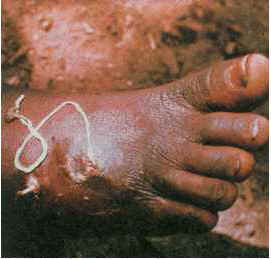
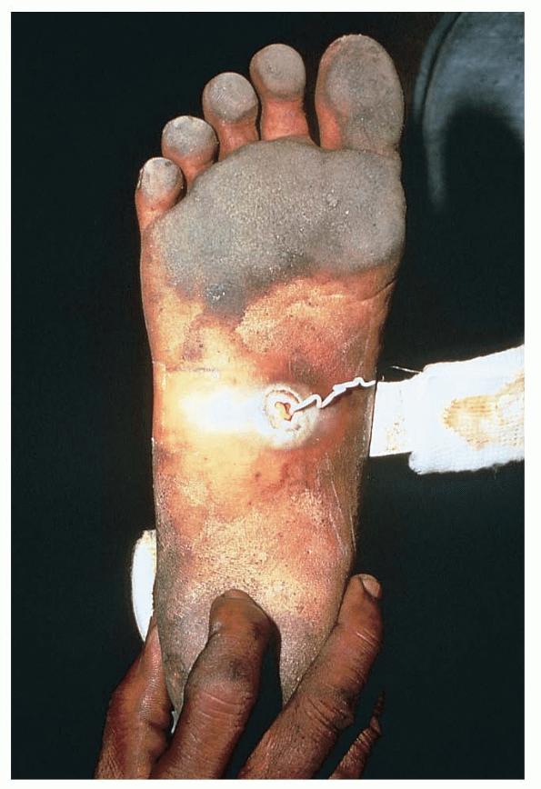
Dracunculiasis Dracunculiasis is an infection caused by the nematode Dracunculus medinensis, also known as the guinea fire worm. D medinensis is in the order Spirurida, an order of parasites that includes the filariae Wuchereria bancrofti, Brugia malayi, and Loa loa. During the last 25 years, concerted efforts to eradicate the guinea worm have been undertaken and these have resulted in a reduction of more than 99% of worldwide cases of dracunculiasis. Current disease incidence is low and is limited specifically to sub-Saharan Africa. The Centers for Disease Control and Prevention (CDC) proposed a global campaign for eradication of dracunculiasis in 1980, and, in 1988, numerous African ministers of health set a target date of 1995 for total eradication. After that target was missed due to slow mobilization in countries with endemic disease, a target date of 2009 was set. Unfortunately, despite considerable progress, that date was also not met. By the end of 2008, dracunculiasis was endemic in 6 countries (Ethiopia, Ghana, Mali, Niger, Nigeria, and Sudan), and the number of cases decreased 52% (from 9,585 in 2007 to 4,619 in 2008).1 Sporadic violence and civil unrest in Sudan and Mali poses the greatest threat to the final eradication of dracunculiasis. The term dracunculus is Latin for "little dragon," a misnomer and reference to the symbol. Thus, when the guinea worm disappears, one of the original inspirations for the discipline of medicine will also disappear. Currently, the infection persists and, although uncommon, can cause significant morbidity.
Ingestion of water that contains infective Dracunculus larvae causes the infection. The larvae reside in an intermediate host, a tiny fresh-water crustacean or copepod of the genus Cyclops. The acidic environment of the stomach and duodenum kills the copepods. The larvae are subsequently released in the stomach or small intestine and penetrate the mucosa to mate and mature in the abdomen or retroperitoneal space approximately 60-90 days after initial infection. The maturation stage can last for up to 1 year, and, during this time, the adult male probably dies because only the female worm is recovered from symptomatic patients. After maturation is complete, the female Dracunculus reaches a length of up to 1 m (with a thickness of only 1-2 mm) and slowly migrates from the GI tract into subcutaneous tissue, usually to a location in the lower extremity. The actual route of migration is unknown. In this subcutaneous location, one or more females prepare larval exit sites through the skin, from whence larvae may be released into another water supply. Free-living larvae can survive only 3 days without a host; they become infective after 2 weeks (2 molts) within the host copepod. Death due to dracunculiasis is not caused by the primary infection and occurs only in cases in which secondary infection of the worm's exit site leads to sepsis . The mortality rate is quite low; however, morbidity is a major concern, with secondary infection being the most common complication. Cellulitis or the formation of an abscess requires prompt attention, and pain from the exit sites often can incapacitate patients for weeks. This is usually observed in individuals who have multiple worms and rely on their ability to stand or walk for their livelihood. Farmers with untreated dracunculiasis in Nigeria have been found to miss work for up to 3 months. Infected schoolchildren may miss up to 25% of the school year. Therefore, Dracunculus infection can cause significant socioeconomic burden for individuals and communities. Another, more chronic, complication of dracunculiasis is encapsulation of the adult worm, which occurs when the calcified remains of the worm persist in the extremity of the patient. This can result in chronic pain and intermittent swelling of the extremity. In a small percentage of individuals who have permanent scarring or deformity of the lower extremity, even after the worm has been extracted, chronic pain may persist for as long as 18 months. Notably, on average, infected individuals have multiple worm extrusions at the same time (1.8 worms per person, on average). Rarely, dracunculiasis can present with worms located in anomalous locations, including the lungs, pancreas, testes, spinal cord, or periorbital tissue.
Causes
The following studies are indicated in dracunculiasis:
Medical Care
Antiparasitic agents
These agents are used to speed the pace of worm extraction. Metronidazole (Flagyl, Protostat)DOC as therapy adjunctive to extraction. Active against various anaerobic bacteria and protozoa. Intermediate-metabolized compounds formed bind DNA and inhibit protein synthesis, causing cell death. Adult250 mg PO tid for 10 d Pediatric25 mg/kg/d PO divided tid for 10 d; not to exceed 750 mg/d Cimetidine may increase toxicity of metronidazole; may increase effects of anticoagulants; may increase toxicity of lithium and phenytoin; disulfiramlike reaction may occur with orally ingested ethanol Documented hypersensitivity; first trimester of pregnancy PregnancyB - Fetal risk not confirmed in studies in humans but has been shown in some studies in animals PrecautionsDo not use during first trimester of pregnancy; use caution with history of hepatic disease or concurrent hepatotoxic drugs; use cautiously with coagulopathies, history of retinal or visual changes, or CNS dysfunction Thiabendazole (Mintezol)Acceptable for use in adults only. Adult50-75 mg/kg/d PO divided bid for 3 d; not to exceed 3 g/d PediatricNot recommended |