| Cow Pox = جدري البقر |
|
|
Cow Pox
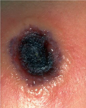
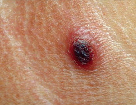
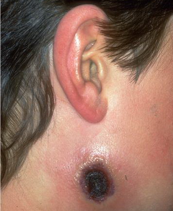
More than 200 years ago, in one of the first demonstrations of vaccination, Edward Jenner inoculated a young English boy with cowpox material from a dairymaid and showed that the boy became resistant to smallpox. Today, cowpox is a rare disease, largely confined to small mammals on the European continent and in Great Britain, with occasional transmission to humans. Most cases present with a small number of vesicopustular lesions on the hands or face that subsequently ulcerate and develop a black eschar before spontaneously resolving. Rarely, cutaneous dissemination and even death may occur. PathophysiologyCowpox is caused by the cowpox or catpox virus, a member of the orthopoxvirus family, which also includes smallpox and vaccinia.1 The virus is believed to be acquired by direct contact with an infected animal, most often a cat in the case of humans, with lesions occurring where the virus gains access through broken skin.2 Infection generally remains localized at the initial site of inoculation, although lymphatic spread in a sporotrichoid pattern and generalized skin infection have been reported.3,4,5 Human-to-human transmission of cowpox has never been reported. As a member of the Orthopoxvirus family, cowpox is a large double-stranded DNA virus that replicates in cell cytoplasm. Viral particles bind to plasma membrane receptors on host cells and then enter into the cytoplasm, where the viral genome is replicated and viral progeny are assembled. After new viral particles are assembled, the host cell lyses, releasing infectious virus, which can enter surrounding cells. Cowpox virus has no latent stage and does not integrate its DNA into the host genome. Poxviruses use numerous strategies to evade the host immune system. These include production of homologues of mammalian tumor necrosis factor receptor, interleukin-1beta receptor, interleukin 18–binding protein, interferon-alpha/beta receptor, and interferon-gamma receptor, as well as a complement-binding protein and a caspase inhibitor.6 These proteins are thought to neutralize the host's antiviral response by binding to cytokines and complement proteins and inhibiting their function. In addition, cowpox virus has been shown to inhibit intracellular transport of major histocompatibility class I molecules, allowing it to evade cytotoxic T cells.7,8
History
Physical
CausesThe natural reservoir of cowpox virus is believed to be small woodland mammals, such as bank voles and wood mice, with humans, cows, and cats being only accidental hosts.
Laboratory Studies
ProceduresSkin biopsy for routine histology, electron microscopy, culture, or molecular detection methods may be performed. Histologic FindingsUsing routine light microscopy, characteristic cytoplasmic inclusions have been observed in biopsies from feline cowpox but not in human material. Immunohistochemistry detects cowpox antigens in feline cases. Using electron microscopy, biopsy material may reveal viral particles.
Medical Care
Surgical Care
Consultations
MedicationBecause most cases of cowpox are mild and self-limited, no treatment is usually required. However, for severe cases with widespread involvement, cidofovir or antivaccinia gammaglobulin may be considered.23 Immune globulinsThe rationale for using antivaccinia gammaglobulin is the presumed cross-reactivity of antibodies to all viruses of the orthopoxvirus family. Antibodies to vaccinia are known to be protective against smallpox and also may be protective against cowpox. Vaccinia immune globulin intravenousRecommended only for severe cases with widespread involvement. This medication and advice on its use may be obtained from the Centers for Disease Control and Prevention Drug Services (404-639-3670). Adult0.6 mL (600 IU)/kg deep IM injection in divided doses over 24-36 h; repeat in 2-3 d if no improvement Pediatric<1 year: 2 mL (2000 IU) IM Increases toxicity of live virus vaccine (MMR); do not administer within 3 mo of vaccine Documented hypersensitivity PregnancyC - Fetal risk revealed in studies in animals but not established or not studied in humans; may use if benefits outweigh risk to fetus PrecautionsCheck serum IgA; infusions may increase serum viscosity and thromboembolic events; infusions may increase risk of migraine attacks, aseptic meningitis (10%), urticaria, pruritus, or petechiae (2-5 d postinfusion to 30 d); increases risk of renal tubular necrosis in elderly and in those with diabetes, volume depletion, and preexisting kidney disease; laboratory result changes associated with infusions include elevated antiviral or antibacterial antibody titers for 1 mo, 6-fold increase in ESR for 2-3 wk, and apparent hyponatremia |