| Cherry hemangiomas = ورم وعائي بشكل الكرز |
|
|
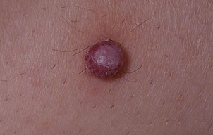
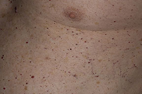
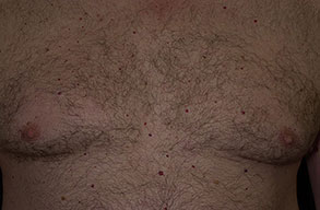
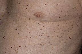
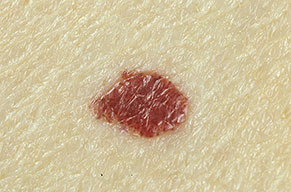
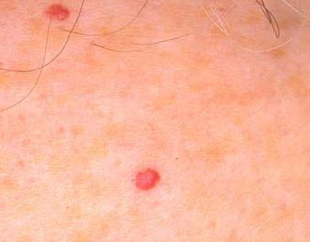
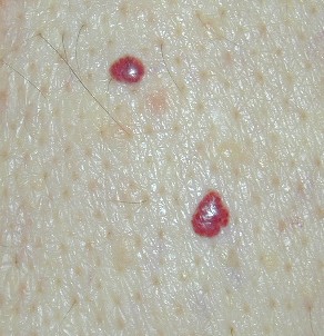
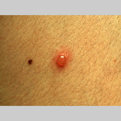
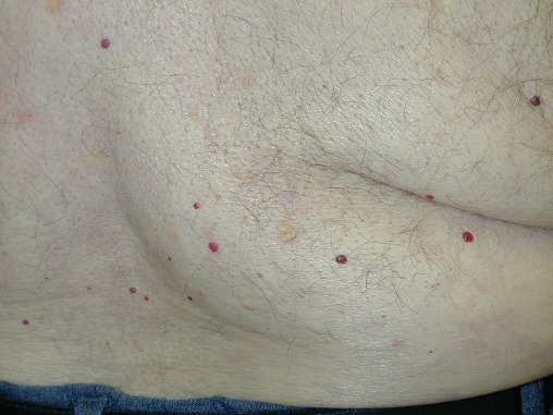
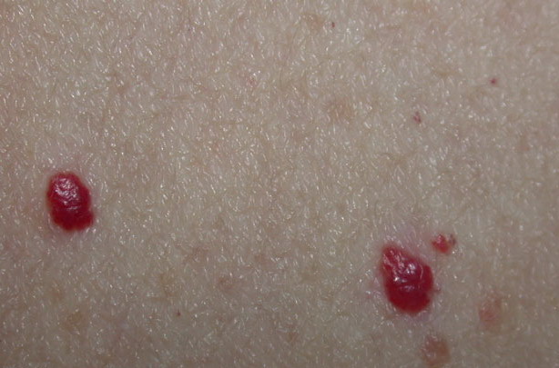
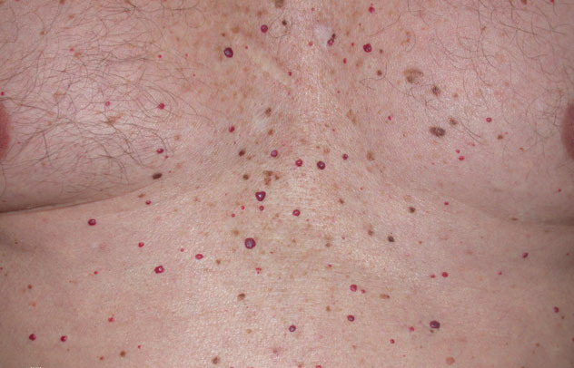
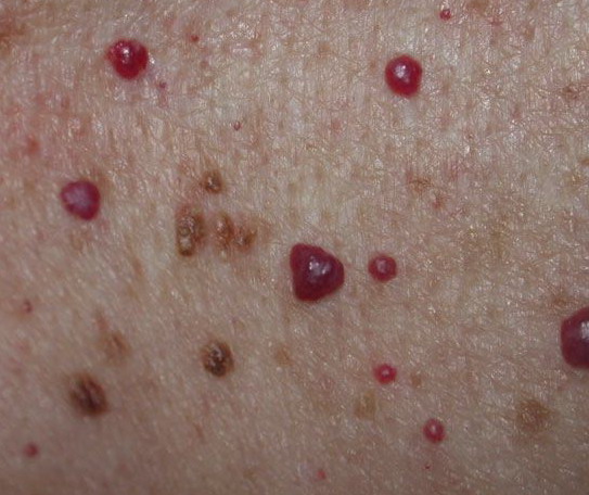
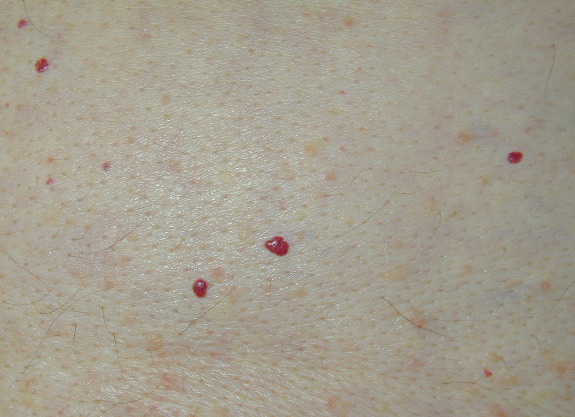
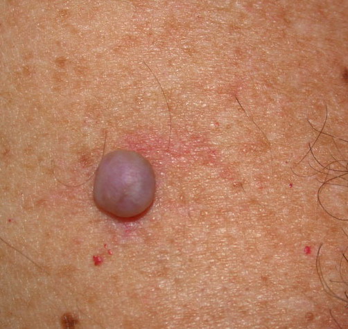
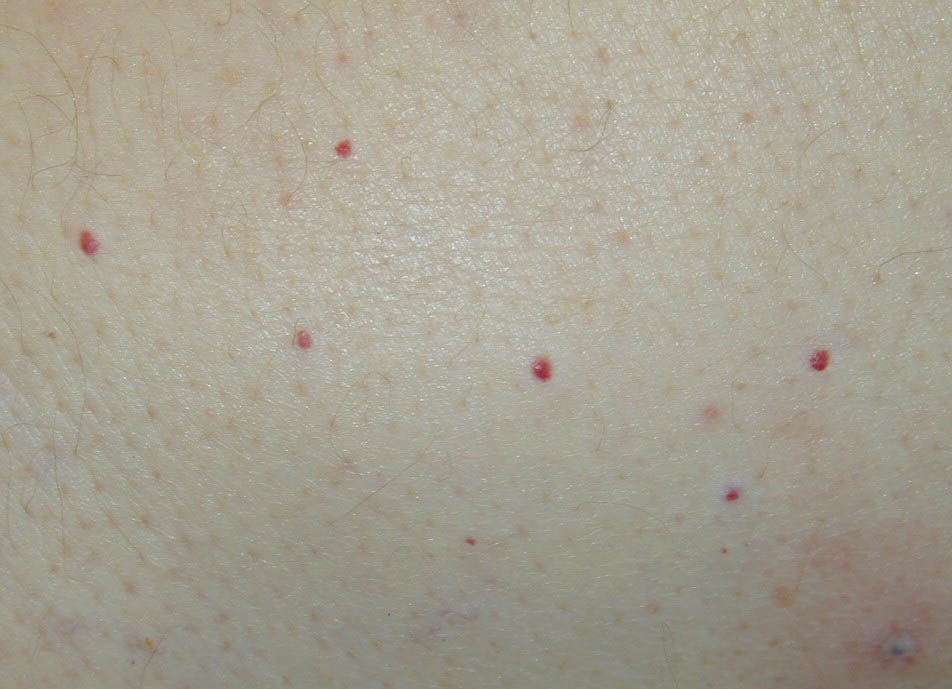
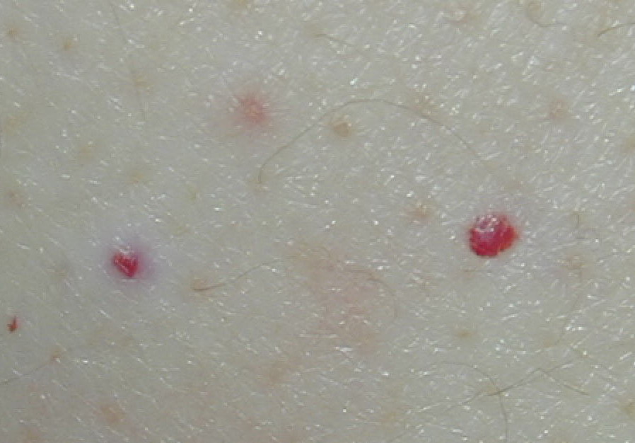
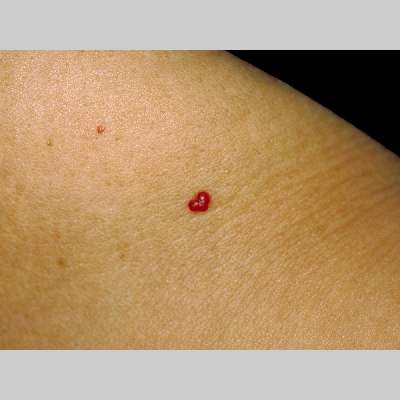
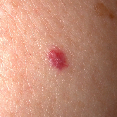
Cherry hemangiomas Cherry hemangiomas are the most common cutaneous vascular proliferations. They are often widespread and appear as tiny cherry red papules or macules.
PathophysiologyInvolvement of cherry hemangiomas is limited to the skin. These benign lesions are formed by a proliferation of dilated venules
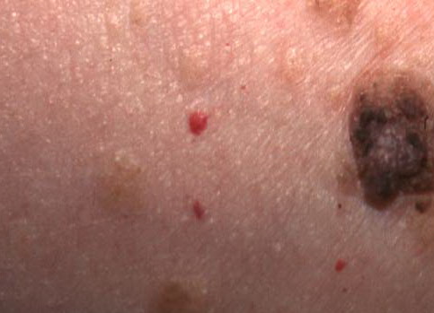
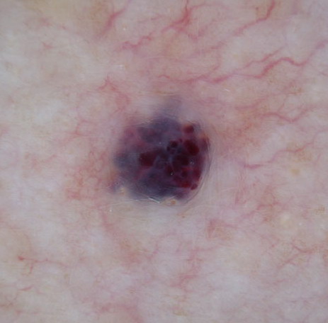
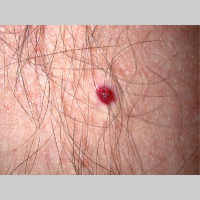
HistoryCherry angiomas typically present in the third or fourth decades of life, and early lesions may appear as small red macules. Lesions may be found on all body sites, but usually, the mucous membranes are spared. Most patients report an increase in number and size of individual lesions with advancing age. PhysicalOn physical examination, lesions may have a variable appearance, ranging from a small red macule to a larger dome-topped or polypoid papule. The color of the lesions typically is described as bright cherry red, but the lesions may appear more violaceous at times . Rarely, a lesion demonstrates a dark brown to an almost black color when a hemorrhagic plug occupies the vascular lumen, often raising concern about the possibility of a malignant melanoma. CausesLittle is known about the factors that contribute to the formation of cherry hemangiomas. Several reports have described the appearance of many small red papules histologically resembling cherry hemangiomas in patients with malignancies,1 although most lesions occur in healthy patients
Laboratory StudiesThe diagnosis of cherry hemangioma is usually made clinically; however, biopsy allows histopathologic confirmation in doubtful situations. ProceduresA skin biopsy (shave or punch) allows histologic confirmation of the diagnosis. Histologic FindingsOn scanning magnification, a sharply circumscribed vascular proliferation usually is noted, often embraced in part by a collarette of epithelium and adnexal structures. Higher magnification demonstrates numerous venules in a thickened papillary dermis. Older lesions often display prominent collagen bundles, which is an appearance suggesting septa. .
Medical CareMedical intervention is not helpful and not indicated in the treatment of the benign vascular proliferations of cherry hemangiomas. Perform biopsy on lesions in which the diagnosis is doubtful. The biopsy procedure may be used as a therapeutic measure to remove traumatized or bleeding lesions. Surgical CareTreatment for cherry hemangioma lesions is recommended only in situations of irritation or hemorrhage or in instances in which the lesions are deemed by the patient to be cosmetically undesirable. Options include the following:
ConsultationsDermatologist consultation may be indicated. For multiple cherry hemangiomas that have appeared over a short period, refer the patient for evaluation to exclude an internal malignancy. In several patients, cherry hemangiomas that have erupted over a very short period of time were associated with an internal malignancy.
Further Outpatient CareIn general, the benign lesions of cherry hemangioma require no therapy, although lesions that are irritated or bleeding (most commonly secondary to trauma) usually require surgical intervention. Follow-up evaluations usually are arranged approximately 1 month after initial therapy. Occasionally, more than a single treatment is required to eliminate the lesion(s). If the lesions are numerous and present as small macules, consider a bleeding disorder such as thrombocytopenia. Deterrence/PreventionNo effective means are available by which the development of the lesions of cherry hemangioma can be prevented. ComplicationsHemorrhages and secondary infection may complicate the course of traumatized lesions, often requiring surgical removal of the inflamed angioma. PrognosisThe appearance of cherry angiomas has essentially no effect on the patient's life span, except in very rare situations in which the angiomas are present as a paraneoplastic sign in association with the development of an internal malignancy.
Rarely, some confusion may arise in determining whether a deeply violaceous or a darkly pigmented papule represents a traumatized and thrombosed cherry angioma or malignant melanoma. In any situation in which doubt exists regarding the diagnosis of a cutaneous neoplasm, perform a skin biopsy and obtain histopathologic analysis. Special ConcernsA 2006 report describes a case in which multiple cherry hemangiomas were noted to occur in a patient while being treated with cutaneous topical nitrogen mustard for vitiligo. The diagnosis of eruptive cherry angiomas was confirmed on histopathology and it was noted that occurrence of new lesions halted when the nitrogen mustard treatment was discontinued |