| Chancroid =القرح اللين |
|
|
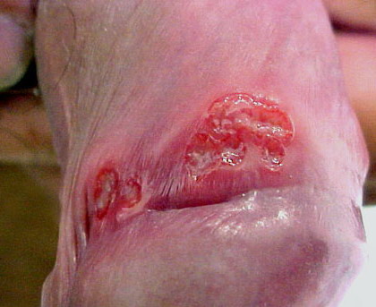
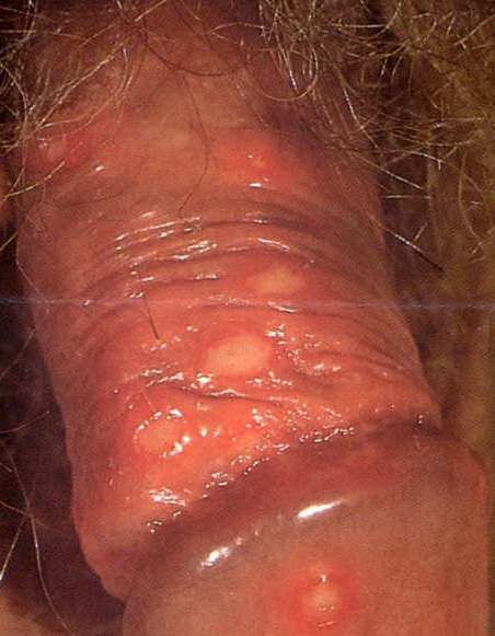
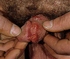
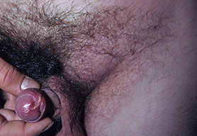
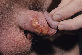
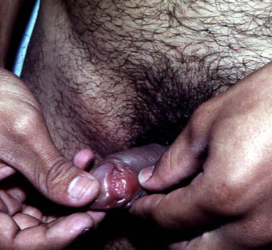
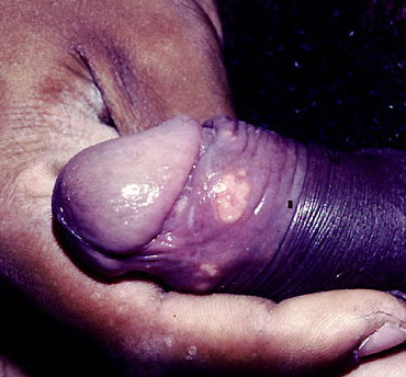
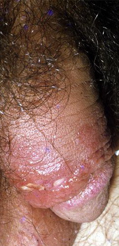
Chancroid ▪ EPIDEMIOLOGY Chancroid is most common in developing countries, especially in Africa and Asia, where the causative organism, Haemophilus ducreyi, was isolated from over 50 percent of genital ulcers in patients until the 1990s.1-3 These endemic regions also have some of the highest rates of human immunodeficiency virus (HIV) infection in the world, and chancroid is common in all 18 countries in which adult HIV prevalence surpasses 8 percent.4 Recent epidemics in the industrialized countries have usually been associated with commercial sex work, the use of crack cocaine, syphilis, and an increased risk of HIV infection.15,16 Lower-class prostitutes appear to be a reservoir in all reported outbreaks of this disease. Men have a markedly higher incidence of chancroid than women.10 Several studies in Africa showed that chancroid ulcer is an important risk factor for the heterosexual spread of HIV-1.17 It is still not clear whether there is an asymptomatic reservoir of H. ducreyi carriers and what the risks of transmission are.18,19 The duration of infectivity in the absence of treatment was estimated to be 45 days for women. The transmission rate from females to males is not known, but a transmission rate from males to females of 70 percent per sex act has been reported.20 ▪ ETIOLOGY AND PATHOGENESIS Historical Aspects H. ducreyi is a Gram-negative, facultative anaerobic coccobacillus that requires hemin (X factor) for growth. The organism is small, nonmotile, and non-sporeforming and shows typically streptobacillary chaining, especially in cultures. The exact taxonomy is still controversial. The current classifications list H. ducreyi as a true Haemophilus sp. However, studies of DNA homology and chemotaxonomy demonstrate substantial differences between H. ducreyi and Haemophilus sp. H. ducreyi will likely be reclassified in the future, but this issue awaits further studies.24,25 Biochemistry Growth Requirements Genetics and Virulence Three major factors seem to be important in the pathogenesis of H. ducreyi infection: the adherence to the epithelial surface, the rate of production of exotoxins (e.g., cytolethal distending toxin),30 and the resistance of the host defense mechanism. Many details about pathogenesis are still unclear. Because of the lack of appropriate experimental model systems, attachment factors, in particular, are not well understood.31 ▪ CLINICAL FINDINGS The incubation period is between 3 and 7 days, rarely more than 10 days. No prodromal symptoms are known. The chancre begins as a soft papule surrounded by erythema. After 24 to 48 hours it becomes pustular, eroded, and ulcerated (Fig. 202-1). Vesicles are not seen. The edges of the ulcers are often ragged and undermined (see eFig. 202-1.1 in on-line edition). The ulcer is usually covered by a necrotic yellowish gray exudate (Fig. 202-2), and its ground is composed of granulation tissue that
CHANCROID AT A GLANCE
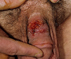
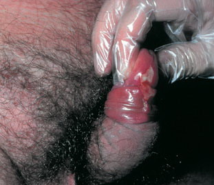
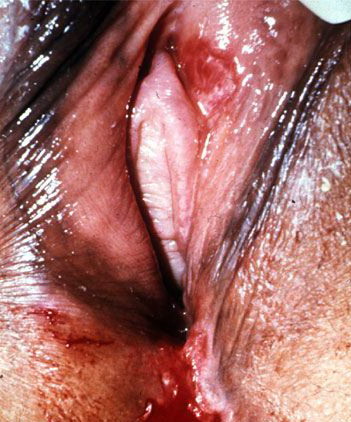
In females the lesions are mostly localized on the vulva (Fig. 202-3), especially on the fourchette, the labia minora, and the vestibule. Vaginal, cervical, and perianal ulcers have also been described. Extragenital lesions of chancroid have been reported on the breasts, fingers, thighs, and inside of the mouth. Trauma and abrasion may be important for such extragenital manifestations. Painful inguinal adenitis (bubo) occurs in up to 50 percent of patients within a few days to 2 weeks (average, 1 week) after onset of the primary lesion (see eFig. 202-3.1 in on-line edition). The adenitis is unilateral in most patients, and erythema of the overlying skin is typical. Buboes can become fluctuant and may rupture spontaneously. The pus of a bubo is usually thick and creamy. Buboes are less common in female patients. In addition to the common types of chancroid described thus far, a number of clinical variants have been reported (Table 202-1). Mild systemic symptoms can accompany chancroid in rare cases, but systemic infection by H. ducreyi has never been observed. Recently chronic skin infection due to H. ducreyi was reported, affecting the lower limb of children visiting Samoa,33 suggesting that this may be a previously unrecognized form of infection.
▪ LABORATORY TESTS Bacterial culture of H. ducreyi currently remains the primary tool for the diagnosis of chancroid in the clinical setting. However, the advent of more sensitive DNA amplification techniques has demonstrated that the sensitivity of H. ducreyi culture is only 75 percent at best.34-36 The bacillus will survive only 2 to 4 hours on a swab unless refrigerated. Swabbed material from the purulent ulcer base should be inoculated directly onto an appropriate culture medium, because no satisfactory transport system is available.37 As noted earlier (see Growth Requirements), the simultaneous use of two primary isolation media from a nutritionally rich agar base supplemented with hemoglobin and serum is recommended for high culture sensitivity.27 Small, nonmucoid, yellow-gray, translucent colonies appear in 2 to 4 days after inoculation. Typically, these colonies remain intact when they are pushed across the agar surface. The identification of H. ducreyi is performed following the recommendations of Lubwama38: demonstration of hemin requirement, oxidase and catalase test, β-lactamase test, and hydrogen sulfide and indole activity. Testing of antibiotic susceptibility is recommended, because clinically significant antimicrobial resistance of H. ducreyi has become common. Direct examination of clinical material by Gram or Giemsa stain may be helpful, but reported sensitivity and specificity values are low—10 percent to 63 percent and 51 percent to 99 percent, respectively.39 The bacilli are usually found in small clusters or parallel chains of two or three organisms streaming along strands of mucus. This pattern has been described as a “school of fish” or “railroad track” (Fig. 202-4). This arrangement, said to be characteristic of H. ducreyi, is nevertheless not pathognomonic, because most genital ulcers have a polymicrobial flora. Cotton or calcium-alginate swabs are recommended for specimen collection. Some authors do not recommend direct microscopy in the routine diagnosis of chancroid.21,36,39 Many attempts have been made to develop serologic tests for chancroid. Due to limited sensitivity in the detection of circulating antibodies to H. ducreyi,
Differential Diagnosis of Chancroid
Polymerase chain reaction (PCR) procedures using different primers have shown greater sensitivity for the diagnosis of chancroid than bacterial culture.39 A multiplex PCR assay has been developed (Roche Molecular Systems, Alameda, CA) for the simultaneous amplification of DNA targets from H. ducreyi, Treponema pallidum, and herpes simplex types 1 and 2,41 which seems to be a particularly attractive diagnostic tool in the investigation of genital ulcers in patients. Multiplex PCR has a resolved sensitivity and specificity for H. ducreyi of 98.4 percent and 99.6 percent, respectively.41 None of these PCR-based methods is commercially available yet, but their use for routine diagnostic purposes would be clearly advantageous. In resource-poor settings in which diagnostic facilities are not readily available, the World Health Organization advocates the use of a syndromic management approach for patients with genital ulcer disease.42 ▪ HISTOLOGY Characteristic histologic features with three vertically arranged zones (superficial necrotic zone, a zone of new blood vessel formation beneath, and a deep zone consisting of dense lymphocytic and plasma cell infiltrate) have been described in chancroid.43 Tissue biopsy is not a recommended diagnostic method, but histologic examination may be useful to exclude malignancy in non-healing or atypical ulcers.39 Staining with H. ducreyi-specific monoclonal antibodies demonstrates the organisms chiefly within the granulocytic infiltrate and fibrin of the ulcer.44 ▪ DIFFERENTIAL DIAGNOSIS (Box 202-1) The three classic causative agents for genital ulceration are H. ducreyi, T. pallidum, and herpes simplex. The clinical appearance of the diseases caused by these three agents can be extremely variable in both men and women, and therefore clinical diagnosis of genital ulcer disease can be made with reasonable certainty for only a minority of patients.45 The cause of genital ulcers46 also differs considerably by geographic region. In industrialized countries isolated painful chancres are most likely due to herpes simplex virus.47 In a high percentage of genital ulcers no pathogen can be isolated, but co-infections of H. ducreyi with T. pallidum (ulcus mixtum) or herpes simplex virus are not uncommon.5,8 ▪ COURSE AND PROGNOSIS The disease is self-limited, and systemic spread does not occur. Occasionally, without treatment, genital ulcer and inguinal abscess have been reported to persist for years. Local pain is the most frequent complaint. If no clinical improvement is evident 1 week after the start of therapy, incorrect diagnosis, co-infection with another organism, concomitant HIV infection, poor compliance with the therapy regimen, or a resistant strain of H. ducreyi must be considered. Infection does not confer immunity, and re-infection is possible. To avoid reinfection, patients must be instructed to use condoms properly.
▪ COMPLICATIONS In about half of untreated patients, the course is that of spontaneous resolution without complications. When treatment is delayed, various complications may occur (Table 202-2). ▪ TREATMENT Based on in vitro susceptibility the most active drugs against H. ducreyi are azithromycin, ceftriaxone, ciprofloxacin, and erythromycin. Worldwide, several isolates with intermediate resistance to either ciprofloxacin or erythromycin have been reported. Regimens actually recommended by the Centers for Disease Control and Prevention, the World Health Organization, and the European Branch of the International Union against Sexually Transmitted Infection are listed in Table 202-3.39,49,50 Antibiotic combinations (e.g., ceftriaxone and streptomycin) showed synergy in an animal model and may be promising to improve singledose treatment, but clinical evaluation is needed.51 Local treatment consists of application of antiseptic dressings (i.e., povidone-iodine). Suppurative nodes should not be incised; if necessary, they can be punctured to prevent spontaneous rupture and sinus tract formation. A large syringe should be used and the fluctuant buboes entered laterally through normal skin. In patients with complicating phimosis a circumcision may be necessary when all active lesions have healed. Even after correct treatment ulcers recur in approximately 5 percent of patients, and retreatment with the original regimen is recommended. Usually reinfection by an untreated sexual partner is the cause of relapse.
HIV infection and lack of circumcision appear to be associated with increased likelihood of infection with H. ducreyi and treatment failure. In resource-poor areas of the world, syndromic management can be recommended, but local epidemiology must be considered. Flowcharts for the management of genital ulcers have been developed that do not require laboratory identification of the causative pathogen. If a patient complains of one or more small blisters or an ulcer with a history of recent blisters, then herpes management should be followed. If an isolated small ulcer and painful matted lymphadenopathy is present, treatment for lymphogranuloma venereum, chancroid, and syphilis should be initiated. If only an ulcer is present, treatment should be for syphilis and chancroid.
|
|||||||||||||||||||||||||||||||||||||||||||||||||||||||||||||||||||||||||||||||||||||||||||||||||||||||||||||||||||||||||||||||||||||||||||||||||||||||||||||||||