Verrucous carcinoma
Verrucous carcinoma (VC) refers to a clinicopathologic concept implying a locally aggressive, clinically exophytic, low-grade, slow-growing, well-differentiated squamous cell carcinoma with minimal metastatic potential.
Verrucous carcinoma typically involves the oral cavity, larynx, genitalia, skin, and esophagus.
In 1948, Ackerman first described verrucous carcinoma in the oral cavity as a low-grade tumor that generally is considered a clinicopathologic variant of squamous cell carcinoma.1 Aird et al first described cutaneous verrucous carcinoma (carcinoma cuniculatum) in 1954.2
Pathophysiology
The pathogenesis of verrucous carcinoma is not yet fully elucidated. Leading theories include human papillomavirus (HPV) infection (anogenital and some oral and sole lesions),3 chemical carcinogenesis induced by smoking and chewing tobacco,4 alcohol consumption and betel nut chewing (oral lesions), and chronic inflammation. Schistosomiasis is associated with verrucous carcinoma of the bladder.
History
The verrucous carcinoma lesion manifests as a verrucous, exophytic, or endophytic mass that typically develops at sites of chronic irritation and inflammation. The verrucous carcinoma tumor enlarges slowly but may penetrate deeply into the skin, fascia, and even bone.5
Physical
Verrucous carcinoma typically involves the oral cavity, larynx, genitalia, skin, and esophagus. Based on the different sites of occurrence, verrucous carcinomas are categorized as follows:
- Oral verrucous carcinoma (Ackerman tumor, oral florid papillomatosis6 )
- The oral cavity is the most common site of occurrence of verrucous carcinoma.7
- Early lesions appear as white, translucent patches on an erythematous base. They may develop in previous areas of leukoplakia, lichen planus,8,9 chronic lupus erythematosus, cheilitis, candidiasis, or submucous fibrosis.
- The more fully developed lesions are white, soft, cauliflowerlike papillomas with a pebbly surface that may extend and coalesce over large areas of the oral mucosa.
- Ulceration, fistulation, and invasion locally into soft tissues and bone (eg, mandible) may occur. Oral verrucous carcinoma most commonly occurs on the buccal mucosa.7 Other sites of involvement are the alveolar ridge, upper and lower gingiva, floor of the mouth, tongue, tonsils, and vermilion border of the lip.
- Painful nonmalignant lymphadenopathy can be seen with concurrent infection or inflammation.
- Tumors most often grow around the lymph nodes rather than metastasizing to them. If metastases do occur, they usually remain limited to the regional lymph nodes.
- Oral verrucous carcinoma involving the hard palate and upper alveolus is considered more aggressive.7
- In these patients, the majority of the tumors present at advanced stages (III-IV).7
- Anogenital-type verrucous carcinoma (Buschke-Löwenstein tumor)
- The Buschke-Löwenstein tumor usually manifests as an exophytic tumor of the genital or perianal area, with ulceration and sometimes fistulae and sinuses.10
- They typically manifest as large, exophytic, nonhealing, cauliflowerlike lesions with a verrucous or ulcerated surface. The Buschke-Löwenstein tumor usually can only be differentiated from ordinary condylomata based on histologic findings. These tumors tend to infiltrate deeply, and recurrence is common.10
- The Buschke-Löwenstein tumor is preferentially seen in men and immunocompromised patients.10 It commonly occurs on the glans penis, mainly in uncircumcised men.
- Less commonly, the Buschke-Löwenstein tumor occurs in the bladder11,12 or on vaginal, cervical, perianal, scrotal, vulvar, and pelvic organs.
- Palmoplantar verrucous carcinoma (epithelioma cuniculatum)13,14,15,16,17
- Most cutaneous verrucous carcinomas are found on the plantar surface of the foot. These tumors most commonly involve the skin overlying the first metatarsal head, but they also occur on the toes, heel, medioplantar region, dorsum, and amputated stumps.18
- Cutaneous verrucous carcinomas have been reported to develop at sites at long-standing cutaneous scars, including gunshot wounds and burn scars.18
- Lesions are usually slow growing, exophytic, and locally invasive, and they can be misdiagnosed for verruca vulgaris.18
- Exophytic tumors with ulceration and sinuses draining foul-smelling discharge cause pain, bleeding, and difficulty walking.
- Palmoplantar verrucous carcinoma is consider to have a very low incidence of metastases.18
- Cutaneous verrucous carcinoma often occurs as a single mass or plaque, but multiple verrucous carcinomas on the feet and ankles have been reported.19
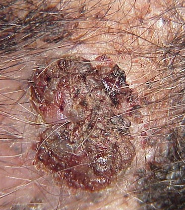
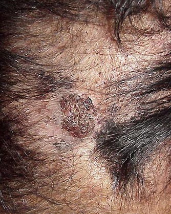
- Verrucous carcinomas arising from other sites (eg, trunk, extremities, scalp, face) also have been reported.
- Others
- Laryngeal verrucous carcinoma usually occurs as a bulky exophytic lesion with a papillomatous appearance that projects from the larynx.20
- Verrucous carcinomas of the endometrium21 and the sinonasal track have been reported.22 Verrucous carcinomas may develop in areas of previous hidradenitis suppurativa23 and genital lichen sclerosus.24
Causes
- HPV infection is thought to facilitate or cause verrucous carcinoma. HPV infection is believed to play the predominate role in the development of verrucous carcinoma of the penis, vulva, and periungual region. HPV-16 has been identified frequently in genital and periungual verrucous carcinoma.25,26 HPV types 6 and 11 are found in the Buschke-Löwenstein tumor.10
- Chronic inflammation may lead to the development of verrucous carcinoma. Inflammatory diseases (eg, long-standing oral ulcerative lichen planus) seem to predispose patients to the development of verrucous carcinoma.
- Associations with verrucous carcinoma have been found in patients who chewed tobacco and betel nuts, dipped snuff, and/or consumed alcohol. Lesions developed at the sites where tobacco was habitually placed in the mouth.7
- Schistosomal infection often is coexistent with verrucous carcinoma of the bladder.
- Oral verrucous carcinoma is associated with poor dental hygiene, ill-fitting dentures, and low socioeconomic status. Oral verrucous carcinoma has a higher incidence in males and in immunocompromised patients
Computed tomography or magnetic resonance imaging may be used to demonstrate the exact location and extent of the verrucous carcinoma (VC) tumor for preoperative staging and surgical planning.
- A skin biopsy always is required for definitive diagnosis of verrucous carcinoma, despite the fact that the diagnosis is suspected strongly on clinical grounds.
- Biopsy is performed routinely in the physician's office using a local anesthetic.
- All skin biopsy specimens obtained to diagnose verrucous carcinoma must reach at least the depth of the mid dermis to allow for determination of the presence or absence of invasive disease.
- A deep (scoop) shave biopsy, a punch biopsy, an incisional biopsy, or an excisional biopsy may be performed.
- Pathology readings preferably are made by a dermatopathologist who has extensive experience with verrucous carcinoma.
Histologic Findings
Verrucous carcinoma of all types may resemble a verruca superficially, with hyperkeratosis, parakeratosis, acanthosis, papillomatosis, and granular cell layer vacuolization. Blunt projections of well-differentiated epithelium surrounded by edematous stroma and chronic inflammatory cells extend into the dermis, sometimes forming sinuses filled with keratin.
Staging
Most verrucous carcinomas are nonmetastatic and are staged based on size, as follows:
- T0 lesions - In situ
- T1 lesions - Less than 2 cm in diameter
- T2 lesions - Between 2 and 4 cm in diameter
- T3 lesions - Greater than 4 cm in diameter
- T4 lesions - Invasive of muscle or bone
Surgical Care
Most physicians treat patients with cutaneous verrucous carcinoma (VC) in their offices. Complete tumor extirpation should be performed at first presentation because verrucous carcinoma can recur, metastasize, and, ultimately, cause death. Recurrent verrucous carcinoma carries a relatively poor prognosis.
Surgical excision and Mohs micrographic (MMS) surgery represent the treatments of choice for cutaneous verrucous carcinomas.
- Excision with conventional margins
- Simple excision is most valuable in the treatment of small verrucous carcinomas of the trunk and extremities and in areas in which tissue sparing is not essential.
- Cure rates following simple excision of well-defined T1 lesions can be as high as 95-99%.
- A 4-mm margin of healthy tissue is recommended for straightforward lesions.
- Standard excision with permanent conventional sections is a highly effective treatment for many verrucous carcinomas. The depth of the excision should include the subcutaneous fat because even small verrucous carcinomas may extend into the subcutaneous fat.
- The disadvantages of excision with an arbitrary margin are that in some cases, the pathology reveals a subclinical positive margin, requiring further surgery. In extensive tumors with inflammatory changes, the surgical margin may be difficult to define. Additionally, more healthy tissue may be excised than is necessary.7
- Mohs surgery27,28,29
- A dermatologic surgeon usually offers MMS. The main advantage of MMS over simple excision in the extirpation of cutaneous verrucous carcinoma is the ability to examine all excision margins (deep and lateral) and to carefully map residual foci of invasive carcinoma.
- MMS provides a cure rate for verrucous carcinoma of 94-100% and has been of particular value in curing verrucous carcinoma with perineural invasion. MMS offers the added benefit of preserving normal tissue, thus facilitating reconstruction.
- MMS is performed routinely in an outpatient setting with the patient under local anesthesia.
- MMS is not widely available outside the United States.
- A multidisciplinary approach using MMS performed in conjunction with a plastic surgeon, otolaryngologist, and radiation oncologist may allow for the complete removal of deeply invasive verrucous carcinoma, preservation of vital structures, and facilitation of the reconstruction of a large operative defect.
- Because of its many advantages, MMS is the procedure of choice for verrucous carcinoma for which tissue preservation is needed. Furthermore, surgery for verrucous carcinoma using MMS may be an integral component in the management of certain verrucous carcinomas that otherwise would be beyond the experience of the cutaneous surgeon.
- Cryosurgery30
- Cryosurgery using liquid nitrogen is a safe and low-cost procedure for the ablation of selected verrucous carcinomas and is well tolerated by patients.
- Cryotherapy has provided a high cure rate for select well-circumscribed superficial verrucous carcinomas. Because of no histologic control, close follow-up is necessary.
- This procedure is the least likely to result in cure and is the least preferred intervention.
- Curettage and electrodesiccation
- Cure rates of 96-99% have been quoted in several large studies for destruction of T0 and T1 verrucous carcinoma (ie, in situ lesions and invasive lesions <2 cm in diameter). This high cure rate was affected by careful patient selection.
- The main disadvantage of curettage and electrodesiccation is a lack of margin control; nonetheless, the procedure is minimally invasive, well tolerated, and effective for in situ lesions without deep involvement.
- Curettage and electrodesiccation is most appropriate for slow-growing lesions of the trunk and extremities.
- Radiation therapy31,32,33
- Radiation therapy offers the potential advantage of avoiding the trauma and deformity of a surgical procedure, but it has occasionally been associated with transformation to high-grade squamous carcinoma.
- Ionizing radiation therapy is used mainly as a treatment for primary cutaneous carcinoma in patients who cannot tolerate surgery (eg, elderly patients).
- Cure rates for T1 lesions range from 85-95%.
- Although the initial cosmetic result following radiation often is good, the long-term result frequently is poor, with atrophy, hypopigmentation, and telangiectasia. Some patients treated with radiation also develop radiation necrosis. This risk increases over time.
- Radiation therapy is not advocated for use over bony structures because of the risk of osteoradionecrosis. Radiation therapy is not advocated for patients who are young or of middle aged.
- Radiation therapy is expensive and requires multiple visits. The procedure is blind to histologic margin control. For these reasons, the use of radiation as primary therapy for verrucous carcinoma generally is restricted to older patients who cannot tolerate or who refuse surgery.
- Other considerations
- Other treatments that have been used for cutaneous verrucous carcinomas with variable success include topical or systemic chemotherapy (bleomycin, 5-fluorouracil, cisplatin, methotrexate), carbon-dioxide laser, intralesional interferon alfa, imiquimod, and photodynamic therapy.34
- In oral verrucous carcinoma in the presence of clinical lymphadenopathy and if the pathological diagnosis is uncertain, neck treatment should be considered
Verrucous carcinoma (VC) usually is cured with appropriate therapy; however, patients at risk for additional verrucous carcinoma and squamous cell carcinoma should be evaluated with a skin examination at 3- to 12-month intervals.
Prognosis
Most patients with verrucous carcinoma have a good prognosis. Local verrucous carcinoma recurrence following definitive treatment is not uncommon. Regarding oral verrucous carcinoma, the reported recurrence rate ranges from 6-40%. Distant metastasis is considered rare. If metastasis does occur, it is mainly at the regional lymph nodes.7 Patients with oral verrucous carcinoma may be at an increased risk of a second primary oral squamous cell carcinoma, which carries a poor prognosis.
|