| Stasis dermatitis =التهاب الجلد الركودي |
|
|
Stasis dermatitis Stasis dermatitis is a common inflammatory skin disease that occurs on the lower extremities in patients with chronic venous insufficiency with venous hypertension. Stasis dermatitis typically affects middle-aged and elderly patients. It rarely occurs before the fifth decade of life, except in patients with acquired venous insufficiency due to surgery, trauma, or thrombosis. Stasis dermatitis is usually the earliest cutaneous sequela of venous insufficiency, and it may be a precursor to more problematic conditions, such as venous leg ulceration and lipodermatosclerosis
Stasis dermatitis occurs as a direct consequence of venous insufficiency. Disturbed function of the 1-way valvular system in the deep venous plexus of the legs results in backflow of blood from the deep venous system to the superficial venous system, with accompanying venous hypertension. This loss of valvular function can result from an age-related decrease in valve competency. Alternatively, specific events, such as deep venous thrombosis, surgery (eg, vein stripping, harvesting of saphenous veins for coronary bypass), or traumatic injury, can severely damage the function of the lower-extremity venous system. The mechanism by which venous hypertension causes the cutaneous inflammation of stasis dermatitis, as shown in the image below, has been extensively studied for decades. Several theories have been proposed. The earliest theories regarding the cause of cutaneous inflammation in venous insufficiency centered on oxygen perfusion of lower-extremity tissues. Originally, an incompetent venous system was thought to lead to pooling of blood in the superficial veins, with reduced flow and therefore reduced oxygen tension in the dermal capillaries. This pooling hypothesis led to the term stasis dermatitis. It was believed that the decreased oxygen content of pooled blood led to hypoxic damage to the overlying skin. The hypoxia/stasis theory was refuted by evidence that instead of pooled, stagnant blood with low oxygen tension, leg veins in patients with venous insufficiency have increased flow rates and high oxygen tension. Arteriovenous shunting could have accounted for these findings, but no evidence of shunting in patients with venous insufficiency was found. The complete lack of evidence to support a hypoxia/stasis theory has led many investigators to advocate the abandonment of the term stasis dermatitis. Subsequent research focused on the role of lower-extremity microcirculation in the pathogenesis of skin damage due to venous insufficiency. In the 1970s and 1980s, increased venous hydrostatic pressure was found to be transmitted to the dermal microcirculation; this leads to increased permeability of dermal capillaries. This increased permeability enables macromolecules, such as fibrinogen, to leak out into the pericapillary tissue; then, polymerization of fibrinogen to fibrin results in the formation of a fibrin cuff around dermal capillaries. It has been hypothesized that this fibrin cuff serves as a barrier to oxygen diffusion, with resulting tissue hypoxia and cell damage. Subsequently, the phenomenon of fibrin cuff formation was found in more severe disease, such as venous ulceration. Fibrin cuffs are not found in ulcers due to causes other than venous hypertension. Decreased cutaneous fibrinolytic activity has been proposed to contribute to the formation of fibrin cuffs.1,2,3 Formation of fibrin cuffs, coupled with decreased fibrinolysis, results in the dermal fibrosis that is the hallmark of advanced stasis dermatitis. Activated leukocytes become trapped in fibrin cuffs and the surrounding perivascular space, releasing inflammatory mediators that contribute to inflammation and fibrosis.4 These leukocytes release the growth factor transforming growth factor-beta1, an important mediator of dermal fibrosis. Furthermore, upregulation of vascular intercellular adhesion molecule-1 (ICAM-1) and vascular cell adhesion molecule-1 (VCAM-1), which are potent chemoattractants to keep leukocytes active in the perivascular environment, occurs.5 The finding of leukocyte-mediated cytokine production, aided by fibrin cuff formation, provides a direct link between dysfunctional venous circulation and cutaneous inflammation with fibrosis.6,7
HistoryPatients with stasis dermatitis typically present with an insidious onset of pruritus affecting one or both lower extremities.
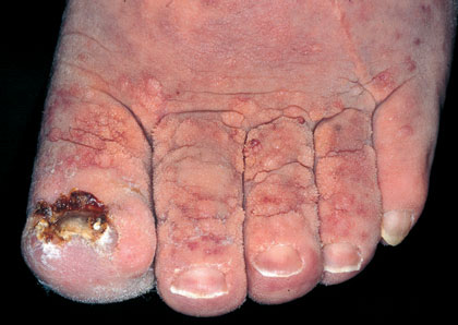
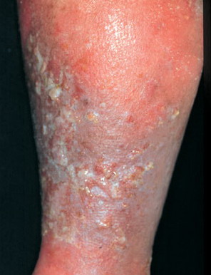
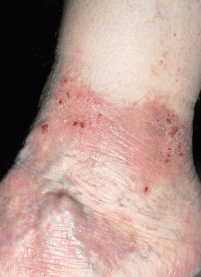
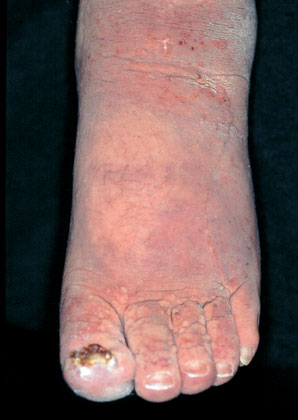
PhysicalPhysical examination in stasis dermatitis patients reveals erythematous, scaling, and eczematous patches affecting the lower extremity, as shown in the images below
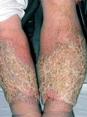
The medial ankle is most frequently and severely involved because of the fact that the medial ankle represents a watershed area with relatively poor blood flow compared with the rest of the leg. In advanced cases of stasis dermatitis, the inflammation may encircle the ankle and extend to just below the knee; this is sometimes referred to as stocking erythroderma. The dorsal part of the foot may be involved in severe cases. A solitary, small patch of stasis dermatitis may mimic basal cell carcinoma or squamous cell carcinoma.12 Involved skin in stasis dermatitis may exhibit the same changes as seen in other eczematous conditions.
Stasis dermatitis frequently occurs along with a background of skin changes that are typical for patients with venous insufficiency. These skin changes include edema, varicosities, hyperpigmentation, atrophic patches (atrophie blanche), and diffuse red-brown discoloration representing deep dermal deposits of hemosiderin (from degraded, extravasated erythrocytes). These chronic changes persist regardless of the activity of stasis dermatitis .
Laboratory StudiesBlood tests are generally not helpful in the management of stasis dermatitis, except in a patient where cellulitis and/or sepsis are suspected. An exception is the patient with stasis dermatitis due to venous thrombosis; patients with venous thrombosis need a thorough hematologic workup to rule out underlying hypercoagulability states. Imaging StudiesRadiologic/Doppler studies may be helpful. In patients with acute new-onset stasis dermatitis or in a young patient, investigating the dynamics of the deep venous circulation is prudent. Venous Doppler studies may reveal deep venous thrombosis or severe valve damage due to past thrombosis. Of course, the consequences of an unrecognized acute or subacute deep venous thrombosis may be catastrophic. Histologic FindingsSkin biopsy of stasis dermatitis, although rarely indicated, shows an acute or subacute dermatitis. Acute lesions may exhibit a superficial perivascular lymphocytic infiltrate, epidermal spongiosis, serous exudate, scale, and crust. Chronic lesions may show epidermal acanthosis with hyperkeratosis. The dermis is characterized by deep dermal aggregates of siderophages due to uptake of hemosiderin from degraded erythrocytes. Dermal capillaries are frequently dilated; long-standing lesions show intimal thickening of small arterioles and venules along with dermal fibrosis. A special consideration in chronic stasis dermatitis where biopsy may be necessary is the development of acroangiodermatitis (pseudo–Kaposi sarcoma). The violaceous plaques and nodules of acroangiodermatitis may be clinically indistinguishable from classic Kaposi sarcoma, especially when occurring in an elderly man. Biopsy samples show changes typical of stasis dermatitis, along with a proliferation of capillaries and fibroblasts. However, the vascular slits and the atypical endothelial cells that are seen in classic Kaposi sarcoma are absent
Compression therapy15 Although extensive work has been completed in the study of treatment of venous ulcers, no large, well-controlled trials examine the treatment of stasis dermatitis. The overall mainstay of treatment has always been aimed at lessening the clinical impact of the underlying venous insufficiency, which is typically accomplished with compression therapy. Assessing the patient's peripheral arterial circulation (clinically or with a Doppler study) before recommending compression therapy is important; adding compression to a leg with compromised arterial circulation could increase claudication and put the patient at risk for ischemic damage. Compression is generally accomplished by means of specialized stockings that deliver a controlled gradient of pressure (measured in mm Hg) to the affected leg. More aggressive compression can be performed by using elastic wraps; compression (Unna) boots; and more sophisticated devices, such as end-diastolic compression boots. Most of these modalities require administration in a physician's office or wound care center. Frequent leg elevation is a necessary adjunct to leg compression. Counseling patients regarding the use of compression therapy is vital to successful management of stasis dermatitis. Although the benefits of compression therapy are widely recognized, noncompliance with compression stockings remain a major barrier to treatment. Difficulty in stocking application, discomfort, and appearance are all reasons for failure to comply with compression therapy.16 Patients frequently resist the idea of compression dressings and/or stockings because these modalities may cause considerable discomfort when first applied to edematous, inflamed lower extremities. However, it is important to reassure patients that this discomfort lessens considerably as leg edema is reduced, and this therapy must be maintained permanently in order to prevent a recurrence of dermatitis and leg ulcers. Compression stockings should be applied early in the morning, before the patient rises from bed, in order to facilitate application when leg edema is at its lowest point. Topical therapy17,18 Topical treatment of stasis dermatitis has much in common with the treatment of other forms of acute eczematous dermatitis. Weeping lesions can be treated with wet-to-damp gauze dressings soaked with water or with a drying agent, such as aluminum acetate. Topical corticosteroids are frequently used for reducing inflammation and itching in acute flares; mid-potency corticosteroids, such as triamcinolone 0.1% ointment, are generally effective. Be wary of the use of high-potency topical corticosteroids in stasis dermatitis because the chronically inflamed skin can increase the risk of systemic absorption and because steroid-induced cutaneous atrophy can predispose the patient to ulceration.19,20 Furthermore, prolonged use of topical steroids can lead to decreased efficacy of the steroid, a phenomenon known as tachyphylaxis. Systemic corticosteroids are not part of stasis dermatitis treatment, although they may be required in very severe cases of widespread autoeczematization. The nonsteroidal calcineurin inhibitors tacrolimus and pimecrolimus may prove to be useful tools in the management of stasis dermatitis. Although these topical medications are approved only for atopic dermatitis, they have been shown to be effective in many steroid-responsive dermatoses. Because the calcineurin inhibitors do not carry the risks of skin atrophy or tachyphylaxis, they have the potential to become valuable agents in the treatment of chronic dermatoses such as stasis dermatitis. Prevention/management of infection Be wary of infection in stasis dermatitis; this becomes more problematic when using topical corticosteroids, which make the patient more susceptible to infection. Open excoriations and erosions should be treated with a topical antibiotic, such as bacitracin or Polysporin. Obvious superficial impetiginization should be treated with topical mupirocin or a systemic antibiotic with activity against Staphylococcus and Streptococcus species (eg, dicloxacillin, cephalexin, cefadroxil, levofloxacin). Culture with sensitivity testing is important when managing suspected superinfection because community-acquired methicillin resistance is becoming increasingly prevalent. Expanded coverage may be necessary in patients who are immunocompromised. Suspected deep cellulitis should always be treated with oral or intravenous antibiotics. Necrotizing fasciitis would be a rare complication but is a surgical emergency. Complications of treatment - Allergic contact dermatitis The development of contact dermatitis is especially problematic in the treatment of patients with stasis dermatitis. Chronic inflammation of the skin, coupled with the use of multiple topical medications (both prescription and over-the-counter) frequently result in contact sensitization as a complication of stasis dermatitis. Patients should be instructed to not apply over-the-counter antibiotics or other topical agents without the direction of a physician. Some of the most frequent contact allergens complicating stasis dermatitis include the topical antibiotics neomycin and bacitracin. In addition, affected patients may become sensitized to rubber products that are found in some wraps and stockings. Topical corticosteroid allergy, while uncommon, is a condition that can worsen stasis dermatitis despite seemingly appropriate prescription therapy. Triclosan-based products have been shown to present a low risk for contact sensitization in patients with stasis dermatitis. These products may be a good alternative to decrease bacterial colonization, especially in patients who have a history of cellulitis or other cutaneous infections.21 Consider contact dermatitis in any patient with stasis dermatitis who becomes clinically worse despite appropriate topical treatment. Long-term management Patients with chronic, quiescent stasis dermatitis can be treated with bland topical emollients to maximize epidermal moisture. Plain white petrolatum is an inexpensive occlusive moisturizer that is very effective and, importantly, does not contain any contact sensitizers. Also see the clinical guideline summary from the Association for the Advancement of Wound Care, Summary algorithm for venous ulcer care with annotations of available evidence. Surgical CareStasis dermatitis related to an arteriovenous fistula or incompetent perforators may respond to ligation of the vessels. Stasis pigmentation, resulting from hemosiderin deposition, is notoriously difficult to treat and typically does not resolve even when the underlying stasis dermatitis is well controlled with topical therapy. However, some authors have reported improvement of stasis pigmentation after treatment with a noncoherent intense pulsed light (IPL) source.22 ConsultationsUncomplicated stasis dermatitis is usually managed in the dermatologist's office.
MedicationRecent new theories regarding the pathogenesis of cutaneous inflammation in venous insufficiency have led to the investigation of systemic therapies, which have been hypothesized to have beneficial modulating effects on neutrophil function. Treatments, such as prostaglandin E1 (PGE1) and pentoxifylline, have been studied in the treatment of venous ulcers; it is hypothesized that these medications decrease cytokine-mediated neutrophil activation, leading to reduced inflammation.23 However, even if these systemic therapies are proven unequivocally effective, it is unlikely that their use will extend beyond the scope of treatment of recalcitrant venous ulcers |