| Purpura = الفرفرية |
|
|
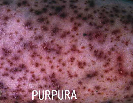
PURPURA
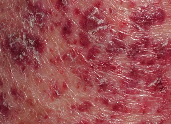
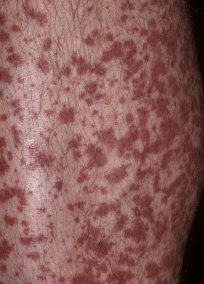
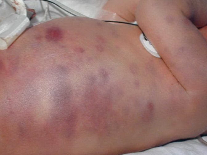
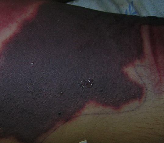
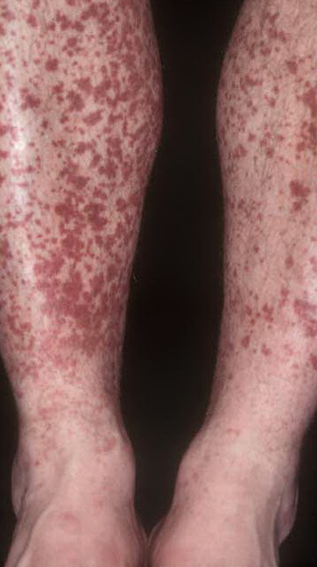
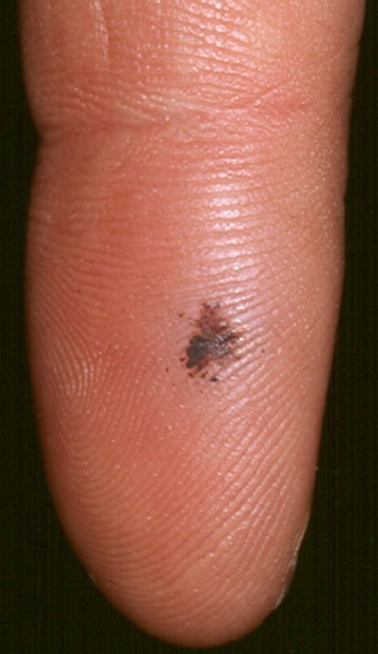
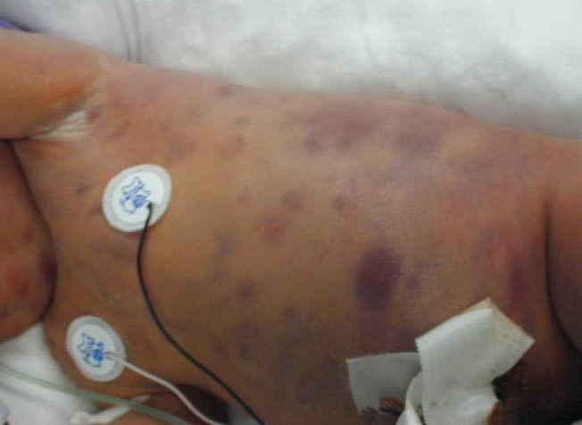
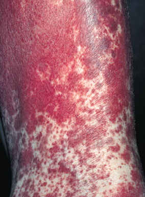
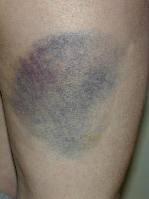
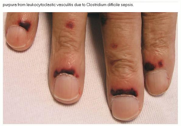
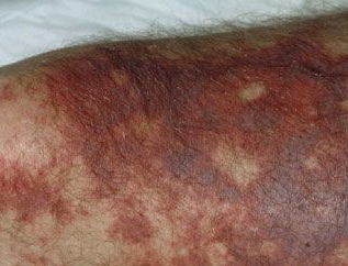
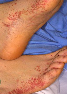
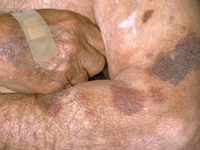
Extravasation of red blood from cutaneous vessels into skin or mucous membranes results in reddishpurple lesions included under the term purpura. The application of pressure with two glass slides or an unbreakable clear lens (diascopy) on a reddish-purple lesion is a simple and reliable method for differentiating redness due to vascular dilatation (erythema) from redness due to extravasated erythrocytes or erythrocyte products (purpura). If the redness is non-blanching under the pressure of the slide, the lesion is purpuric. Petechiae are small, pinpoint purpuric macules. Ecchymoses are larger, bruise-like purpuric patches. These lesions correspond to a noninflammatory extravasation of blood. As extravasated red blood cells decompose over time, the color of purpuric lesions change from bluish-red to yellowish-brown or green. If a lesion is purpuric and palpable (“palpable purpura”), the suggestion of an inflammatory insult to the vessel wall as a cause of extravasation of blood and inflammatory cells exists. A clinical example is leukocytoclastic vasculitis
|