Mycetoma Maduromycosis
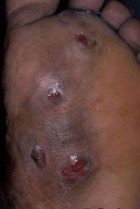
Madura Foot
Mycetoma is a chronic localized infection caused by different species of fungi or actinomycetes. It is characterized by the formation of aggregates of the causative organisms, known as grains, that are found within abscesses. These either drain via sinuses onto the skin surface or involve adjacent bone, causing a form of osteomyelitis. Grains are discharged onto the skin surface via these sinuses. The disease advances by direct spread, and distant metastatic sites of infection are very rare. Mycetomas caused by species of fungi are known as eumycetomas, and those caused by aerobic actinomycetes or filamentous bacteria are known as actinomycetomas . The organisms are usually soil or plant saprophytes that are only incidental human pathogens.
EPIDEMIOLOGY
Mycetomas are mainly, but not exclusively, found in the dry tropics where there is low annual rainfall They are sporadic infections that are seldom common, even in endemic areas. Occasionally, nonimported cases are reported from temperate climates, although in these cases, the most common organism is Scedosporium apiospermum. Actinomycetomas due to Nocardia sp. are most common in Central America and Mexico. In other parts of the world, the most common organism is a fungus, Madurella mycetomatis. The actinomycete Streptomyces somaliensis is isolated most often from patients originating from Sudan and the Middle East. The causative organisms of mycetoma have been isolated from either soil or plant material, including Acacia thorns, in endemic areas.
The organisms are implanted subcutaneously, usually after a penetrating injury. It is unusual to find any underlying predisposition in patients with mycetoma, and the persistence of the organism after the initial inoculation appears to be related to its ability to evade host defenses through a variety of adaptations such as cell wall thickening and melanin deposition.
CLINICAL FINDINGS
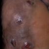
The clinical features of both fungal and actinomycete mycetomas are very similar. They are most common on the foot, lower leg, or hand, although head or back involvement also may occur. Infection of the chest wall is most characteristic of Nocardia infections . The earliest stage of infection is a firm, painless nodule that spreads slowly with the development of papules and draining sinus tracts over the surface . Local tissue swelling, chronic sinus formation, and later bone involvement distort and deform the original site of infection . Lesions are seldom painful except in the late stages and where sinus tracts are about to emerge onto the skin surface. Dissemination from the initial site is exceptionally rare, although local lymphadenopathy may occur.
X-ray changes include periosteal erosion and proliferation, as well as the development of lytic lesions in the bone. Bone scans or magnetic resonance imaging may identify bone lesions at an earlier stage.
DIFFERENTIAL DIAGNOSIS
Chronic bacterial or tuberculous osteomyelitis may resemble
mycetoma. Actinomycosis is also similar but usually develops close to certain sites, such as the mouth or the cecum, where the causative organisms are sometimes commensal.
LABORATORY TESTS
Finding the mycetoma grains is the key to establishing the diagnosis, and these are generally discharged from the openings of sinus tracts. However, they also may be obtained by removing the surface crust from a pustule or sinus tract with a sterile needle and gently squeezing the edges. Grains are 250- to 1000-µm white, black, or red particles that can be picked out with the naked eye . Direct microscopy of grains is important because it will show whether the grain is composed of the small actinomycete or broader fungal filaments. In general, it is not possible to distinguish the fine actinomycete filaments in potassium hydroxide (KOH) mounts or, for that matter, in hematoxylin and eosin-stained material. In addition, black grains are always caused by fungi; red grains, by actinomycetes .
Final identification requires isolation of the causal agent in culture. In view of the number of possible species, a series of different culture media and conditions of incubation should be used. Morphologic and physiologic characteristics are used to distinguish between the genera and species. There are now a few examples where the organism has been identified using specific primers through use of the polymerase chain reaction. Serology is diagnostically helpful only in some cases (e.g., in S. somaliensis), and even then, more as a guide to therapeutic response.
Histologically, there is a chronic inflammatory reaction with neutrophil abscesses and scattered giant cells and fibrosis. Grains (50 to 250 µm) are found in the center of the inflammation. Their size and shape may help in the identification, although with non-pigmented eumycetomas, this is seldom sufficient .
TREATMENT
Of the fungal causes of mycetoma, some cases of M. mycetomatis infection respond to ketoconazole, 200 mg daily, over several months. For the others, a trial of therapy with griseofulvin, terbinafine, or itraconazole is worth attempting. However, responses to chemotherapy are unpredictable, although antifungals may slow the course of infection. Surgery, usually amputation, is the definitive procedure and may have to be used in advanced cases. However, the value of major surgery in a disfiguring but nonlife-threatening infection has to be weighed against the availability of appropriate prosthetic limbs.
Actinomycetomas generally respond to antibiotics such as a combination of dapsone with streptomycin or sulfamethoxazole-trimethoprim plus rifampin or streptomycin. Amikacin also may be used in recalcitrant Nocardia infections. The responses in all but a few cases are good.