


LAMELLAR ICHTHYOSIS
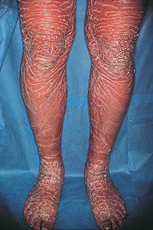
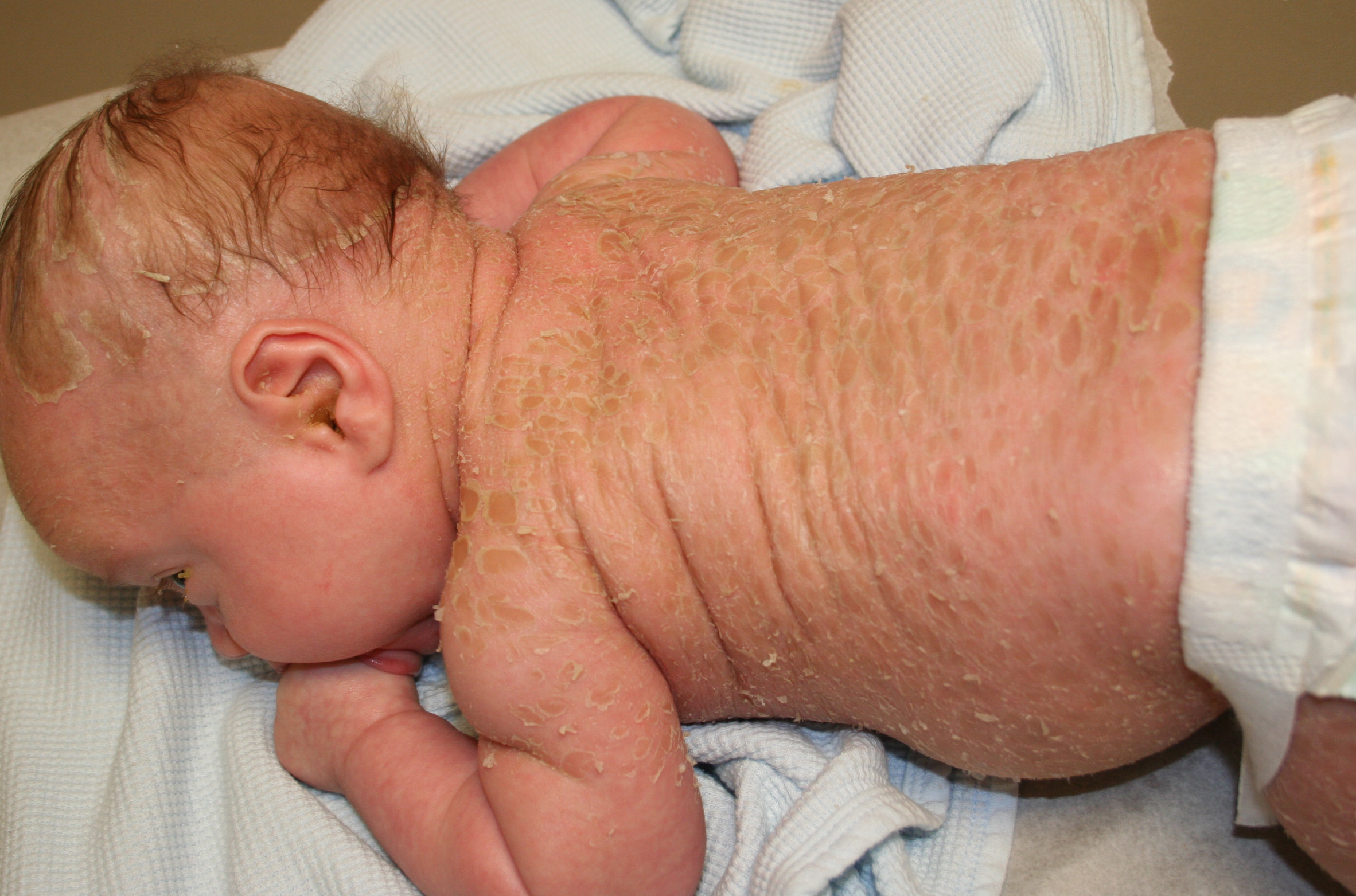
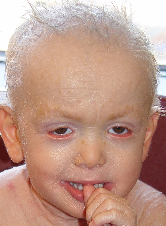
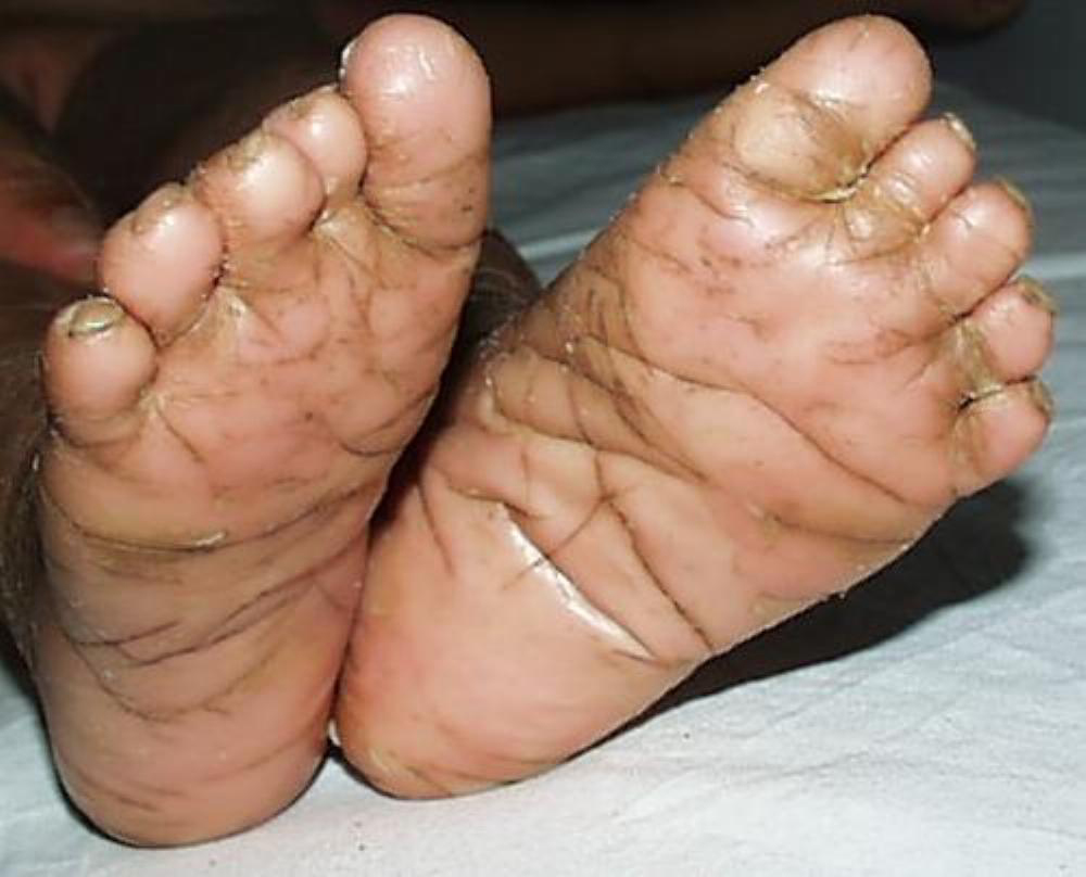
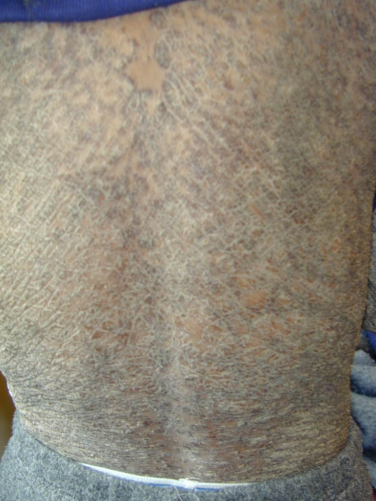
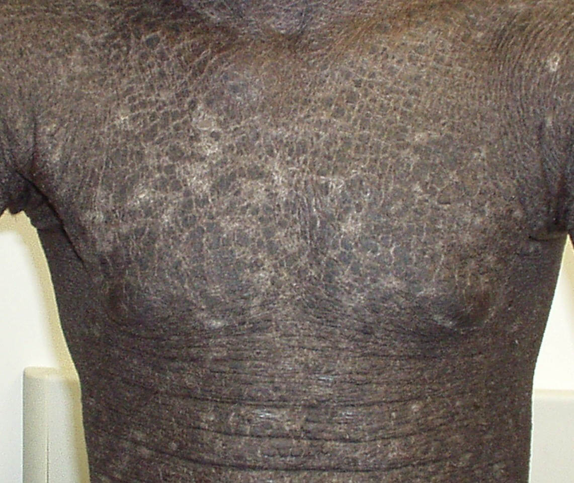
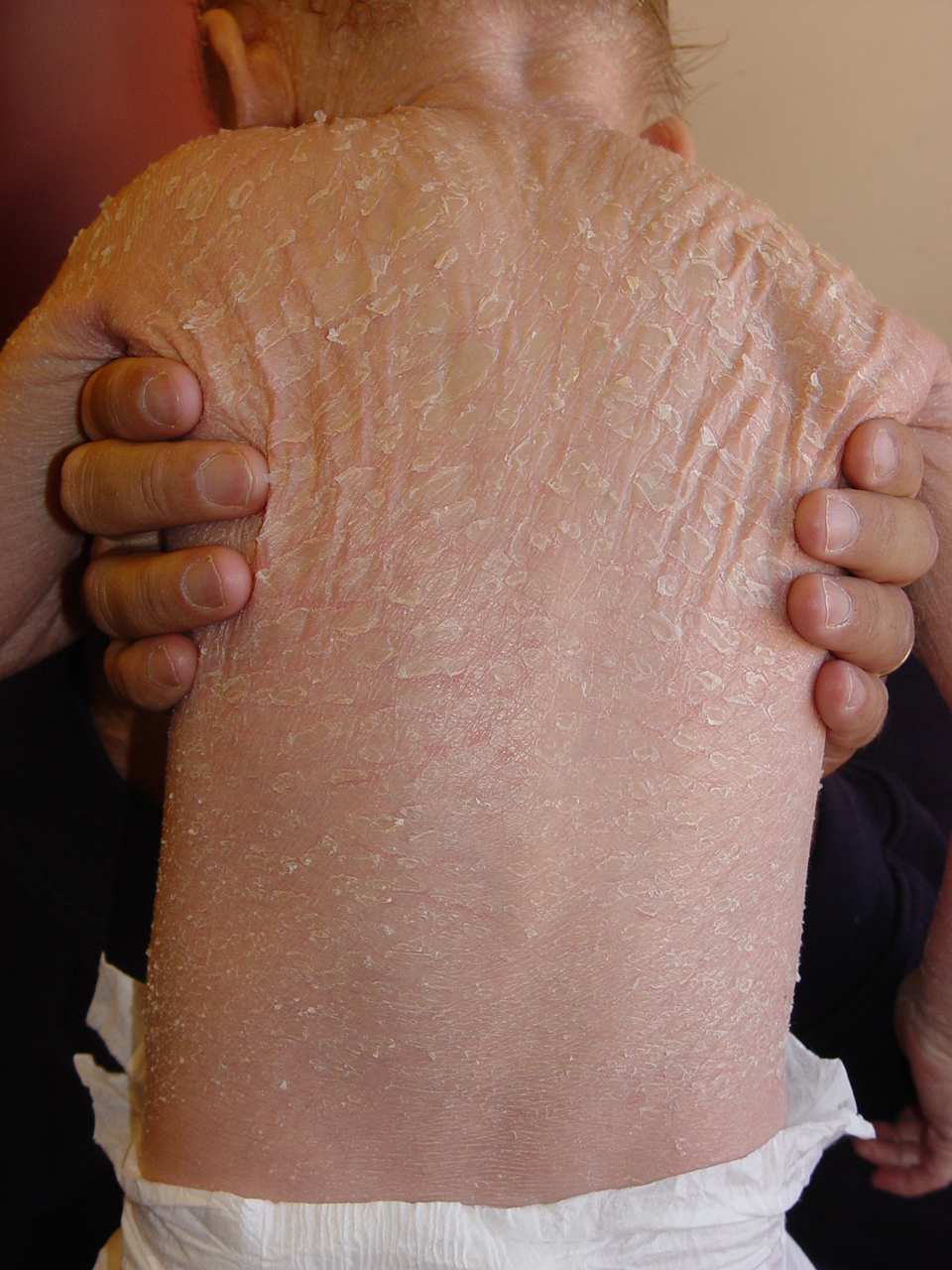
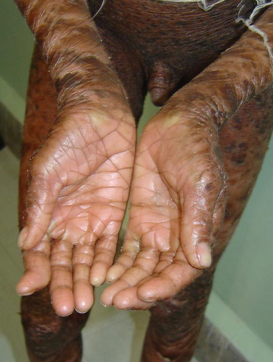
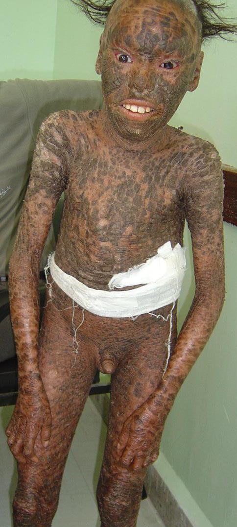
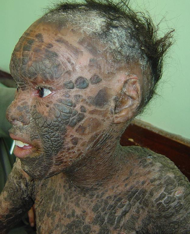
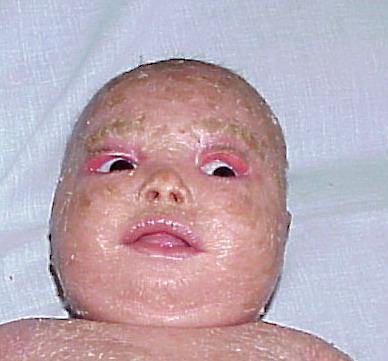
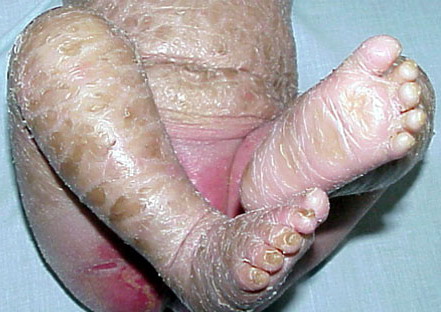
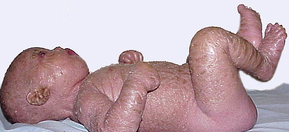
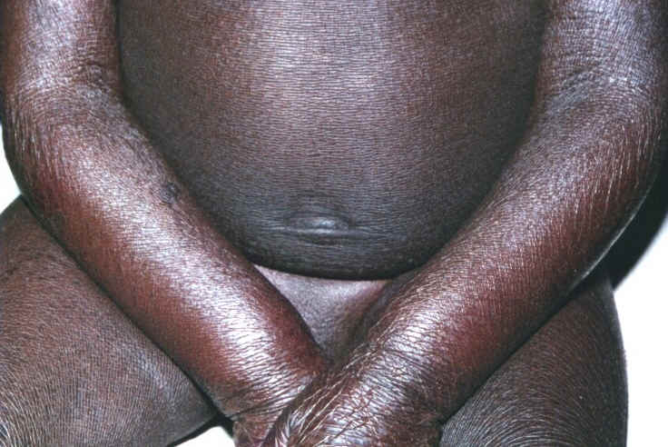
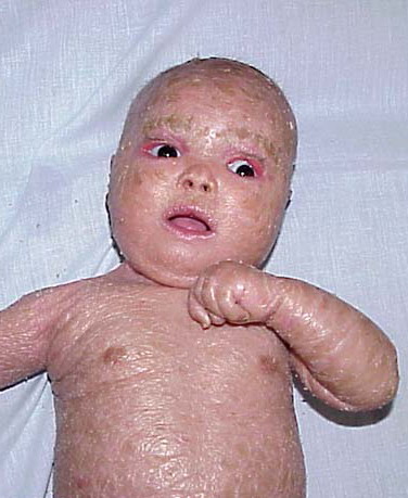
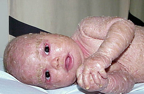
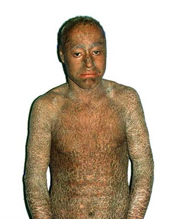
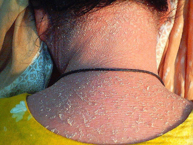
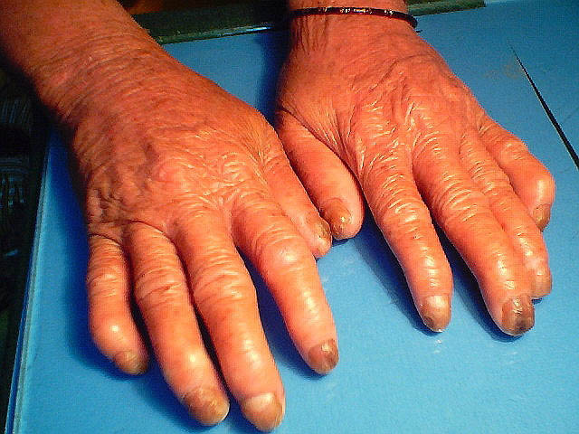
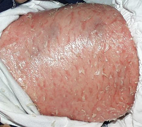
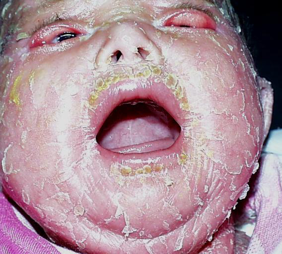
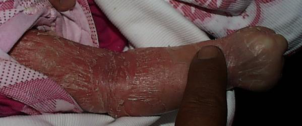
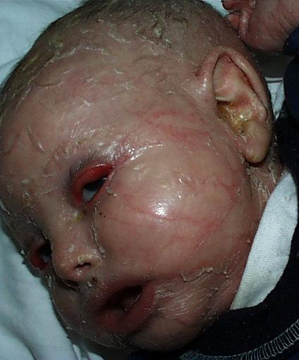
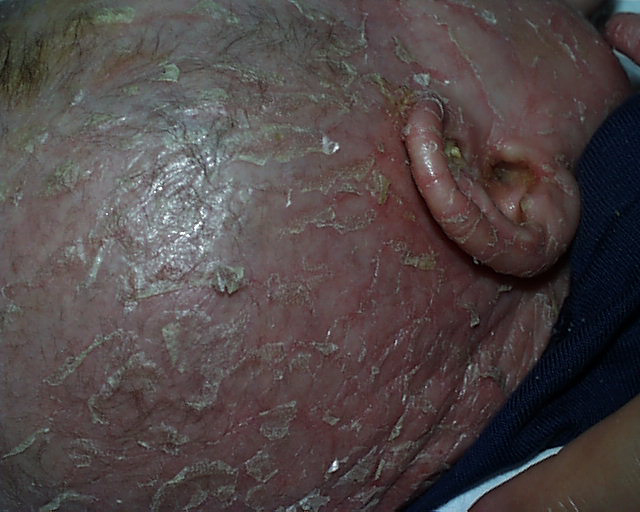
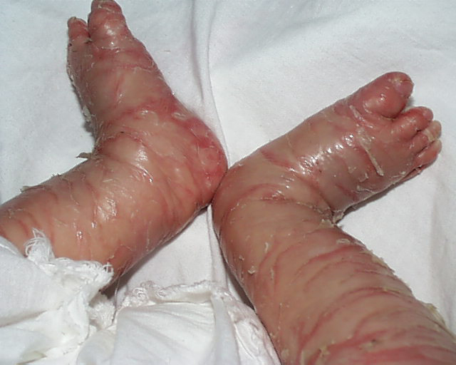
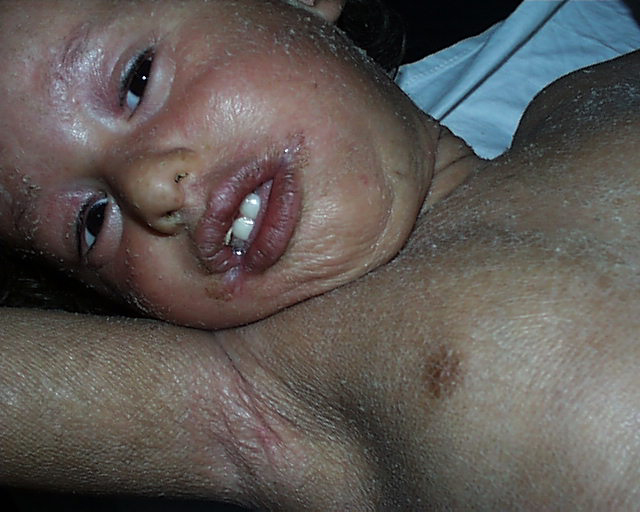
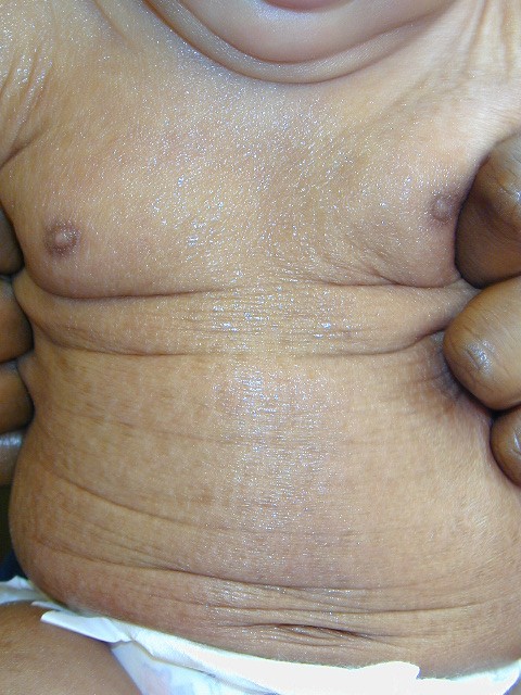
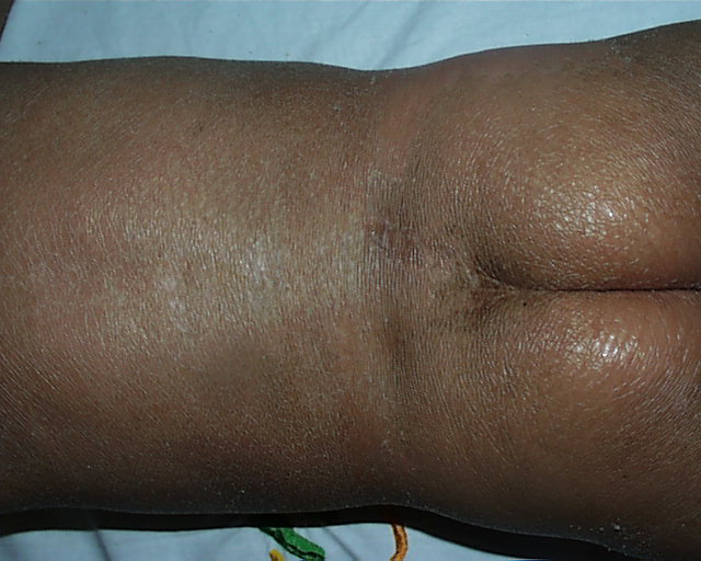
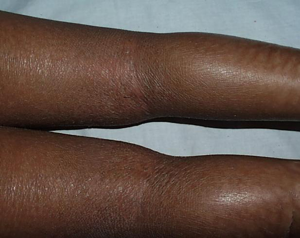
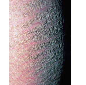
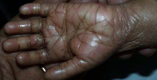
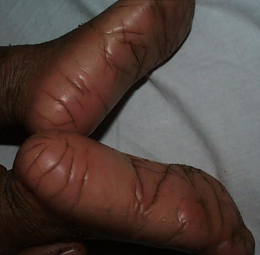
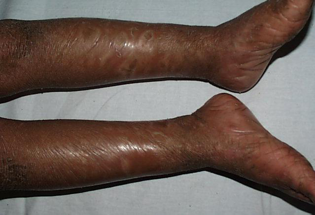
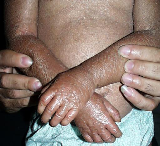
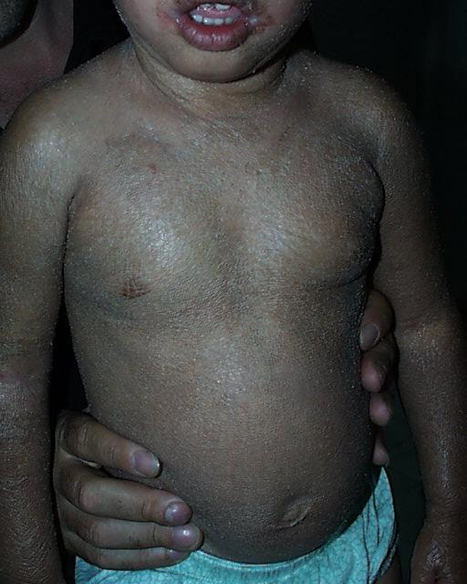
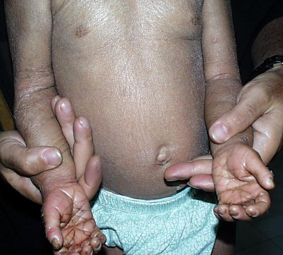
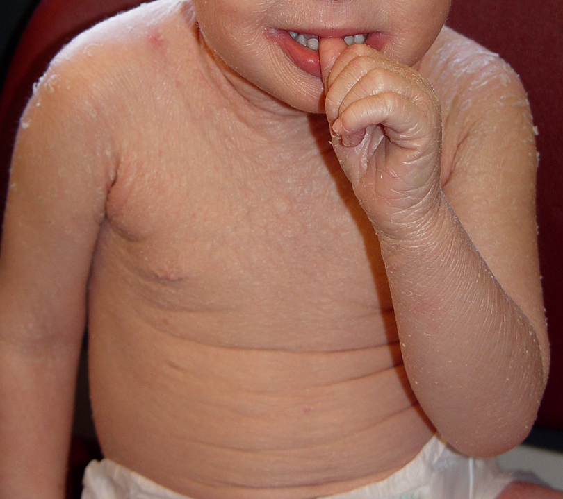
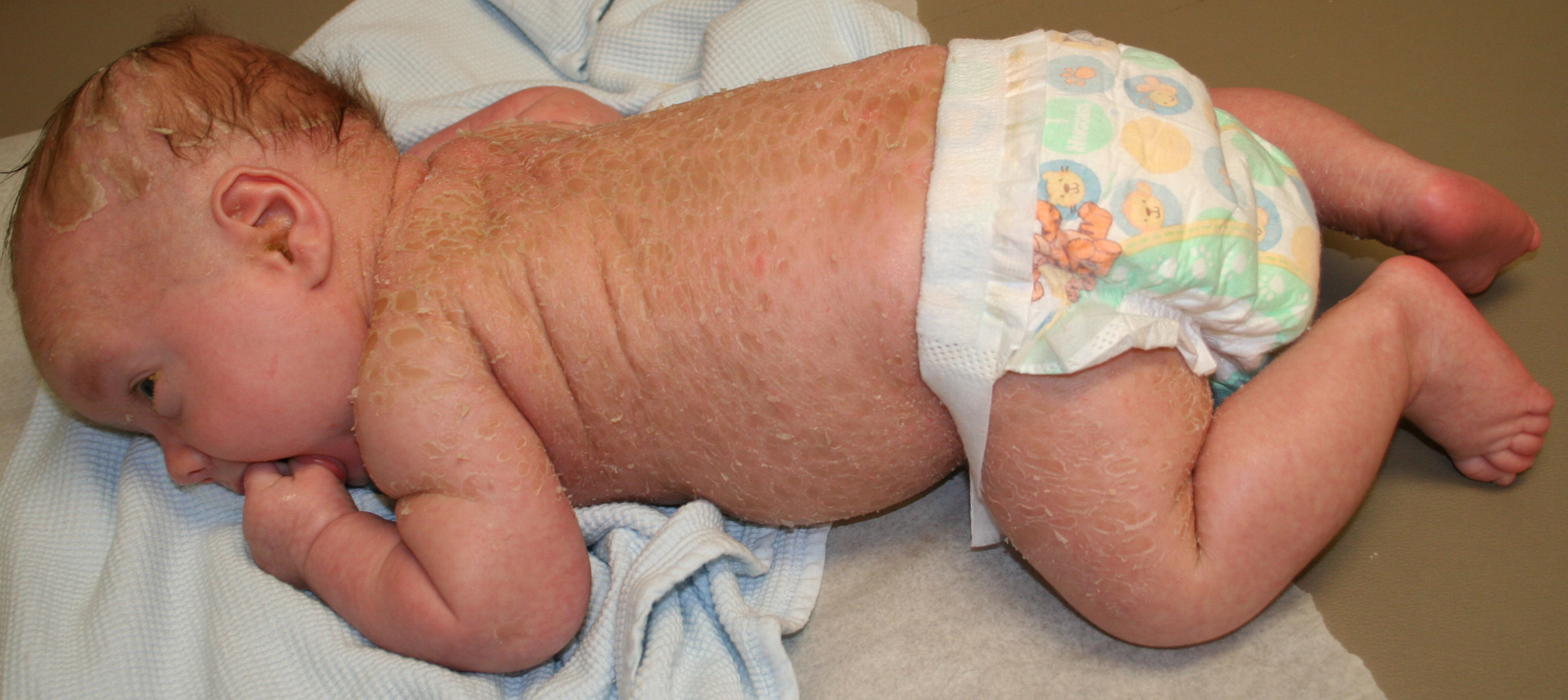
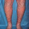
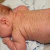
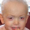
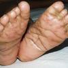
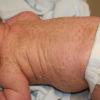
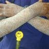
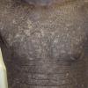
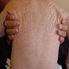
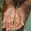
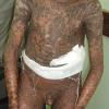
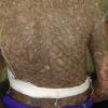
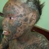
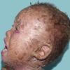
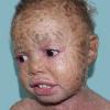
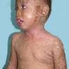
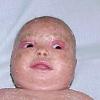
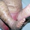
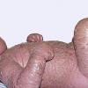
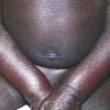
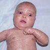
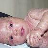
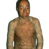
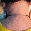
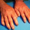
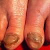
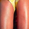
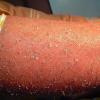
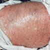
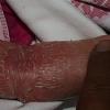
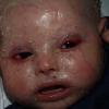
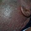
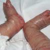
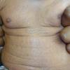
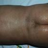
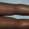
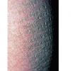
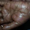
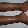
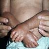
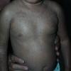
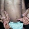
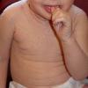
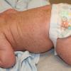
Lamellar ichthyosis is apparent at birth, and the newborn usually presents encased in a collodion membrane. At this time, the skin may be red. Over time, the skin develops large, plate-like scales, which appear to be arranged in a mosaic pattern . In some areas, the scales are centrally attached with raised borders. The scales tend to be largest over the lower extremities, where the large, plate-like scale separated by superficial fissuring can lead to an appearance similar to that of a dry riverbed. During childhood and into adulthood the degree of erythema may vary, but the severe presentation of classic lamellar ichthyosis usually has minimal to no erythroderma. Involvement of the palms and soles in lamellar ichthyosis is variable and ranges from minimal hyperlinearity to severe keratoderma.
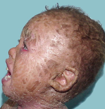
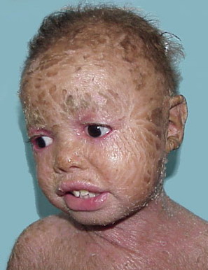
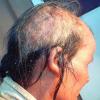
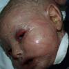
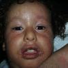
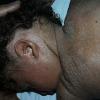
The lips and mucous membranes tend to be spared in lamellar ichthyosis, but the adnexal structures may be compromised by the adherent, firm scale. Thick stratum corneum on the scalp tends to encase hairs, and in conjunction with the tautness of the skin, may lead to a scarring alopecia, most marked at the periphery of the scalp. The hyperkeratosis can interfere with normal sweat gland function, resulting in hypohidrosis, but the degree of impairment varies between patients. Some patients have severe heat intolerance and must be vigilant to avoid overheating. Treatment with oral retinoids can improve or prevent some sequelae of lamellar ichthyosis. Patients frequently notice an increase in sweating, with improved heat tolerance. Although retinoid therapy can cause blepharitis or even conjunctivitis, it is usually well tolerated by patients with lamellar ichthyosis. Moreover, the ability of systemic retinoid (and in some cases, topical retinoid) therapy to decrease thick periocular scale can decrease the tendency to develop ectropion. Nevertheless, patients with severe, classic lamellar ichthyosis usually require careful eye maintenance. Because of the ectropion , the lids may fail to close fully, particularly during sleep; hydration with liquid tears during the day and ophthalmic lubricants at night can prevent exposure keratitis.
Histopathologic examination typically shows orthokeratotic hyperkeratosis with
mild to moderate acanthosis . Rates of epidermal proliferation are normal or only slightly elevated in patients with lamellar ichthyosis, in contrast to those with CIE, which has significantly greater labeling indices.
|
Genes Identified in Autosomal Recessive Congenital Ichthyosis
|
|
GENEa
|
PROTEIN
|
FUNCTION
|
SYSTEMIC FINDINGS
|
|
TGM1
|
Transglutaminase 1
|
Cornified envelope formation
|
—
|
|
ABCA12
|
ATP-binding cassette, subfamily A, member 12
|
Membrane transport/lipid metabolism
|
—
|
|
ALOXE3
|
Arachidonate lipoxygenase 3
|
Hydroperoxide isomerase
|
—
|
|
ALOX12B
|
Arachidonate 12-lipoxygenase, R type
|
Lipoxygenase
|
—
|
|
ICHYN
|
Ichthyin
|
Trioxilin A3 receptor? Hepoxylin pathway?
|
—
|
|
FLJ39501 (CYP4F22)
|
Cytochrome P450, family 4, subfamily F, polypeptide 2
|
LTB4-omega-hydoxylase?
|
—
|
|
SNAP29
|
SNAP receptor (SNARE)
|
Vesicle transport
|
CEDNIK syndrome
|
|
ABHD5 (CGI58)
|
Abhydrolase domain containing 5 comparative gene identification-58
|
Member of esterase/lipase/thioesterase subfamily
|
Chanarin-Dorfman syndrome
|
|
ATP = adenosine triphosphate; CEDNIK = cerebral dysgenesis, neuropathy, ichthyosis, and palmoplantar keratoderma; LTB4 = leukotriene B4; SNAP = soluble N-ethylmaleimide-sensitive factor attachment protein.
|
|
a The same gene may have more than one name.
|
|
Genetic linkage studies performed on a group of families affected with the severe phenotype of classic lamellar ichthyosis (large, plate-like scale; ectropion; and minimal to no erythema) found linkage to markers on chromosome 14 in the region of the transglutaminase 1 gene locus. Subsequently, mutations in TGM1, the gene encoding transglutaminase 1, were found in several families with lamellar ichthyosis, solidifying the role for transglutaminase 1 in the formation of a normal stratum corneum and its role as a cause of lamellar ichthyosis. The transglutaminases catalyze calcium-dependent cross-linking of proteins through the formation of ε-(γ-glutamyl) lysine isodipeptide bonds. During the formation of the stratum corneum, transglutaminase catalyzes the cross-linking of cellular proteins, mostly involucrin, loricrin, and small proline-rich proteins. The resulting protein complex is deposited on the inner side of the plasma membrane to form the cornified envelope. Transglutaminase also attaches ceramides to cornified envelope proteins, notably involucrin, and thereby is important in the formation of both the protein and lipid components of the stratum corneum.
Bathing suit ichthyosis is an unusual form of lamellar ichthyosis in which affected individuals develop the scaling typical of lamellar ichthyosis, but limited to the bathing suit area. The distribution correlates with warmer areas of skin. Decreased transglutaminase is found in these areas, and unique, temperature-sensitive mutations in TGM1 have been identified in affected individuals.
In a human skin/immunodeficient mouse xenograph model, transfer of a transglutaminase 1 gene into transglutaminase 1-deficient keratinocytes from lamellar ichthyosis patients resulted in normalization of transglutaminase expression and epidermal architecture, in addition to restoration of cutaneous barrier function. Additional disease-causing loci have been found .