| Acute cutaneous lupus erythematosus=الذئبة الحمامية الجلدية الحادة |
|
|
Acute cutaneous lupus erythematosus
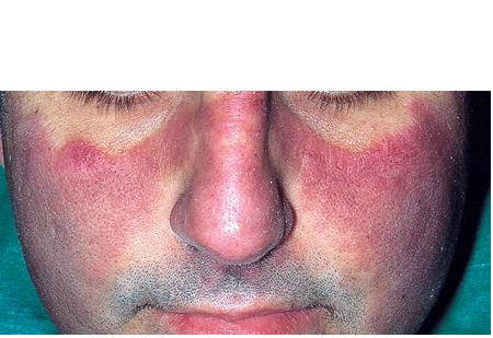
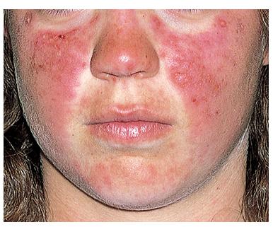
Lupus erythematosus is a heterogeneous connective-tissue disease associated with polyclonal B-cell activation and is believed to result from the interplay of genetic, environmental, and hormonal factors. The spectrum of disease involvement can vary from limited cutaneous involvement to devastating systemic disease. From a dermatologic standpoint, the type of skin involvement can prove to be a good barometer of the pattern of underlying systemic activity. Lupus erythematosus–specific skin diseases are recognized in 3 categories, including (1) acute cutaneous lupus erythematosus (ACLE), (2) subacute cutaneous lupus erythematosus (SCLE), and (3) chronic cutaneous lupus erythematosus (CCLE). Clinical characteristics of each group are unique, although histopathologically, only subtle differences are identified. The focus of this article is acute cutaneous lupus erythematosus. Acute cutaneous lupus erythematosus refers to a typical malar eruption in a butterfly pattern localized to the central portion of the face and/or a more generalized maculopapular eruption representing a photosensitive dermatitis. Acute cutaneous lupus erythematosus has a strong association with the systemic disease for which patients present to rheumatologists and internists.
PathophysiologyThe etiology of lupus erythematosus is believed to be multifactorial, involving genetic, environmental, and hormonal factors. An association with human leukocyte antigen DR2 and human leukocyte antigen DR3 has been identified. Concordance in monozygotic twins and familial associations support a genetic basis in acute cutaneous lupus erythematosus. Certain viruses (eg, Epstein-Barr virus, cytomegalovirus, HIV) have been implicated in precipitating or exacerbating lupus erythematosus. Chemicals such as L-canavanine, which is present in alfalfa sprouts, have been known to induce systemic lupus erythematosus (SLE)–like illness. Drugs implicated in inducing a lupus erythematosus–like illness (eg, procainamide, isoniazid, hydralazine) are uncommonly associated with cutaneous manifestations.
Immunopathology Data concerning direct immunofluorescence in acute cutaneous lupus erythematosus are sparse. In one study, the results of 5 (100%) of 5 skin biopsy specimens were reported as positive for the lupus band test. The lupus band test refers to the presence of immunoglobulins and C3 complement components along the dermoepidermal junction. All 3 immunoglobulin classes (immunoglobulin G [IgG], immunoglobulin M [IgM], immunoglobulin A [IgA]) and a variety of complement components have been identified at the dermoepidermal junction. Research has shown that 60% of patients with a malar eruption of lupus erythematosus have positive lupus band test results. In nonlesional skin, positive lupus band test results correlate strongly with an aggressive course of systemic disease.3
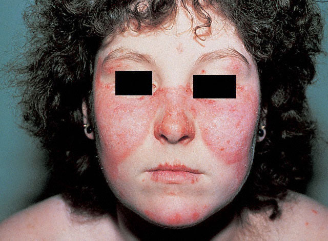
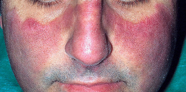
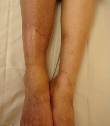
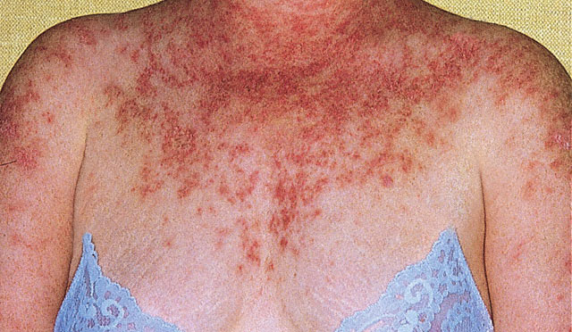
HistoryThe most common presentation of acute cutaneous lupus erythematosus is a red macular eruption involving the malar area. The forehead, periorbital area, and neck also may be involved, representing a photodistribution. Occasionally, unilateral involvement may occur, as shown below Less commonly, acute cutaneous lupus erythematosus presents as a generalized photosensitive eruption, while more rarely, patients present with widespread blistering simulating toxic epidermal necrolysis (TEN). TEN is believed to be a phototoxic reaction .
Acute cutaneous lupus erythematosus can be transient, lasting for several days to weeks. Lesions wax and wane with sun exposure over a period of several hours; however, some patients experience prolonged disease activity. Resolution of lesions may result in postinflammatory hyperpigmentation, especially in patients with darkly pigmented skin. Usually, the lesions are nonscarring. Patients with acute cutaneous lupus erythematosus frequently experience superficial ulceration of the oral and nasal mucosae. These lesions may produce extreme discomfort in some patients, although the lesions may be entirely painless in others. The posterior surface of the hard palate is the site affected most frequently; however, the gingival, buccal, and lingual mucosae also may be involved. Note that acute cutaneous lupus erythematosus may coexist with other lupus erythematosus–specific skin diseases. Localized acute cutaneous lupus erythematosus lesions have been observed in 20% of subacute cutaneous lupus erythematosus patients; however, occurrence of acute cutaneous lupus erythematosus with chronic cutaneous lupus erythematosus is unusual. Physical
CausesIn patients who are disposed genetically to developing systemic lupus erythematosus, the disease can be triggered by viruses (eg, EBV) and exposure to ultraviolet light. Medications typically do not induce acute cutaneous lupus erythematosus in patients with drug-induced lupus erythematosus
Laboratory Studies
Procedures
Histologic FindingsThe most striking change in acute cutaneous lupus erythematosus is the presence of edema involving upper dermis and focal liquefactive degeneration of the basal cell layer. Cellular dermal infiltrate is sparse and consists of lymphocytes. In extreme cases, dissolution of the basal layer occurs secondary to extensive vacuolization, forming a subepidermal bulla
Medical Care
Consultations
DietDietary restrictions may be necessary in the presence of renal compromise. ActivityAdvise patients to avoid activities involving excessive exposure to the sun. MedicationThe goals of pharmacotherapy are to reduce morbidity and to prevent complications. CorticosteroidsHave anti-inflammatory properties and cause profound and varied metabolic effects. Corticosteroids modify the body's immune response to diverse stimuli. Prednisone (Sterapred)Glucocorticoid (adrenocortical steroid) absorbed easily into GI tract. Immunosuppressant for treatment of autoimmune disorders; may decrease inflammation by reversing increased capillary permeability and suppressing PMN activity. Stabilizes lysosomal membranes and also suppresses lymphocytes and antibody production. Adult0.5-1 mg/kg/d PO prn for short periods PediatricAdminister as in adults Ketoconazole, erythromycin, clarithromycin, estrogens, birth control pills increase levels; aminoglutethimide, phenytoin, phenobarbital, rifampin, cholestyramine, and ephedrine decrease levels Absolute: Systemic fungal infection; herpes simplex keratitis; hypersensitivity (usually with corticotropin, occasionally with IV preparations) PregnancyB - Fetal risk not confirmed in studies in humans but has been shown in some studies in animals PrecautionsAbrupt discontinuation of glucocorticoids may cause adrenal crisis; hyperglycemia, edema, osteonecrosis, myopathy, peptic ulcer disease, hypokalemia, osteoporosis, euphoria, psychosis, myasthenia gravis, growth suppression, and infections may occur. ImmunosuppressivesUsed for immunosuppression and, ultimately, for disease control. Azathioprine (Imuran)Antagonizes purine metabolism and inhibits synthesis of DNA, RNA, and proteins. May decrease proliferation of immune cells, which results in lower autoimmune activity. For dermatomyositis/polymyositis, respiratory and muscular symptoms respond but skin lesion response has not been consistent. Adult1 mg/kg/d qd or bid (empiric) or by TPMT level (see Precautions); increase dose by 0.5 mg/kg/d after 6-8 wk prn; increase q4wk, not to exceed 2 mg/kg/d for most dermatologic purposes PediatricInitial: 2-5 mg/kg/d PO/IV Allopurinol increases risk of pancytopenia; captopril/ACE inhibitors may increase risk of anemia and leukopenia; increased dose of warfarin may be necessary; may need increased dose of pancuronium for adequate paralysis; live-virus vaccines, co-trimoxazole (increased risk of hematologic toxicity); rifampicin (transplants possibly rejected); clozapine (increased risk of agranulocytosis) Absolute: Documented hypersensitivity, pregnancy or attempting pregnancy, and clinically significant active infection PregnancyD - Fetal risk shown in humans; use only if benefits outweigh risk to fetus PrecautionsTPMT testing not entirely reliable; it involves testing TPMT activity in RBCs, which correlates with systemic TPMT activity; functional enzyme test shown to have variability between test sites, and kits may contain varying amounts of enzyme inhibitor; starting at low doses, monitoring for pancytopenia, and then increasing dose is alternative; if clinical response is not good, patient may be a homozygote for high activity and may need an increased dose Cyclophosphamide (Cytoxan, Neosar)Chemically related to nitrogen mustards. As an alkylating agent, the mechanism of action of the active metabolites may involve cross-linking of DNA, which may interfere with growth of normal and neoplastic cells. Adult500-750 mg/m2 IV qmo PediatricAdminister as in adults Allopurinol may increase risk of bleeding or infection and enhance myelosuppressive effects; may potentiate doxorubicin-induced cardiotoxicity; may reduce digoxin serum levels and antimicrobial effects of quinolones; chloramphenicol may increase half-life while decreasing metabolite concentrations; may increase effect of anticoagulants; coadministration with high doses of phenobarbital may increase rate of metabolism and leukopenic activity; thiazide diuretics may prolong cyclophosphamide-induced leukopenia and neuromuscular blockade by inhibiting cholinesterase activity Documented hypersensitivity; severely depressed bone marrow function PregnancyD - Fetal risk shown in humans; use only if benefits outweigh risk to fetus PrecautionsRegularly examine hematologic profile (particularly neutrophils and platelets) to monitor for hematopoietic suppression; regularly examine urine for RBCs, which may precede hemorrhagic cystitis Thalidomide (Thalomid)Immunomodulatory agent that may suppress excessive production of tumor necrosis factor-alpha and may down-regulate selected cell-surface adhesion molecules involved in leukocyte migration. Adult100-300 mg/d PO qd with water, preferably hs and at least 1 h pc PediatricNot established May increase sedation of alcohol, barbiturates, chlorpromazine, and reserpine; women must use 2 additional methods of contraception or abstain from intercourse because of teratogenic effects Documented hypersensitivity PregnancyX - Contraindicated; benefit does not outweigh risk PrecautionsPerform pregnancy test within 24-h period prior to initiating therapy (weekly during first month, followed by monthly tests in women with regular menstrual cycles or q2wk with irregular menstrual cycles); bradycardia may occur; use protective measures (eg, sunscreens, protective clothing) against exposure to sunlight or UV light (eg, tanning beds); prescribing physician must register with STEPS provider registry established by manufacturer Hydroxychloroquine (Plaquenil)Inhibits chemotaxis of eosinophils, locomotion of neutrophils, and impairs complement-dependent antigen-antibody reactions. Adult310 mg PO qd or bid for several wk depending on response; 155-310 mg/d for prolonged maintenance therapy Pediatric3-5 mg base/kg/d PO qd or divided bid; not to exceed 7 mg/kg/d Serum levels increase with cimetidine; magnesium trisilicate may decrease absorption; may increase digoxin levels; do not give with chloroquine due to increased retinal toxicity Absolute: Hypersensitivity, retinopathy from any cause PregnancyC - Fetal risk revealed in studies in animals but not established or not studied in humans; may use if benefits outweigh risk to fetus PrecautionsCaution in hepatic disease, G-6-PD deficiency, psoriasis, and porphyria; not recommended for long-term use in children; perform periodic (6 mo) ophthalmologic examinations; test periodically for muscle weakness Immune globulin IV (Sandoglobulin, Gammagard, Gamimune, Gammar-P)Neutralize circulating myelin antibodies through anti-idiotypic antibodies; down-regulates proinflammatory cytokines, including INF-gamma; blocks Fc receptors on macrophages; suppresses inducer T and B cells and augments suppressor T cells; blocks complement cascade; promotes remyelination; may increase CSF IgG (10%). Adult2g/kg IV over 10-12 h PediatricNot established Antibodies in globulin preparation may interfere with response to live viral vaccines (eg, MMR); defer using live viral vaccines until approximately 11 mo after immunoglobulin administration; no known drug interactions No absolute contraindication other than documented hypersensitivity; patients who are IgA deficient should receive IVIG preparations with no IgA; anti-IgE/IgG antibodies, severe thrombocytopenia, or coagulation disorders PregnancyC - Fetal risk revealed in studies in animals but not established or not studied in humans; may use if benefits outweigh risk to fetus PrecautionsCheck serum IgA level before IVIG (use an IgA-depleted product, eg, Gammagard S/D); infusions may increase serum viscosity and thromboembolic events; infusions may increase risk of migraine attacks, aseptic meningitis (10%), urticaria, pruritus, or petechiae (2-30 d postinfusion); increases risk of renal tubular necrosis in elderly patients and in patients with diabetes, volume depletion, and preexisting kidney disease; laboratory result changes associated with infusions include elevated antiviral or antibacterial antibody titers for 1 mo, 6-fold increase in ESR for 2-3 wk, and apparent hyponatremia Monoclonal AntibodyRituximab (Rituxan)Murine/human chimeric anti-CD20 monoclonal antibody. CD20 is expressed early in pre-B cell development. Binding induces complement-dependent B-cell cytotoxicity along with antibody-dependent cellular toxicity. Adult375 mg/m2 qwk for 4 wk (usual dose); no standardized regimen established; early open-label phase I/II study showed varying doses, from 1 infusion of 100 mg/m2 to 4 weekly infusions of 375 mg/m2, without cyclophosphamide bolus or glucocorticoid treatment, significantly reduced Systemic Lupus Activity Measure score over 1 y in 65% of patients PediatricNot established . |