Acquired acral fibrokeratoma
In 1968, Bart et al1 described 10 cases of an uncommon acquired growth that was located on the fingers. Although it clinically resembled a cutaneous horn or rudimentary supernumerary digit, it had distinct histopathological findings. The authors named this growth acquired digital fibrokeratoma (ADFK). Subsequently, Pinkus2 reported 28 more cases; however, because the lesions Pinkus described also occurred on the proximal hand, toes, soles, and one in the prepatellar region, he suggested the entity might be more appropriately called acquired acral fibrokeratoma.
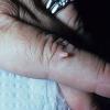
An acquired periungual fibrokeratoma is similar to an acquired acral fibrokeratoma, differing primarily in that the former arises from the proximal nail fold. Koenen tumors, although similar, occur in association with tuberous sclerosis and histologically may have atypical stellate myofibroblasts.3
Pathophysiology
Despite the fact that most patients deny a history of precedent trauma, the major hypothesis is that subclinical injury contributes to the development of acquired digital fibrokeratomas.
History
Most acquired digital fibrokeratoma patients present with an asymptomatic protuberance.
Physical
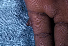
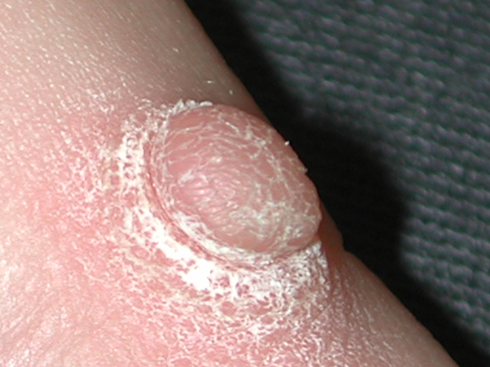
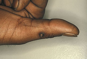
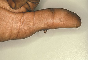
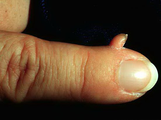
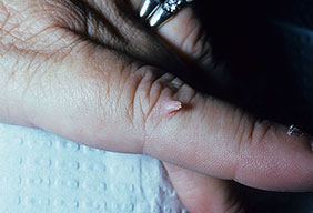
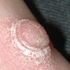
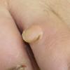
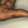
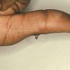
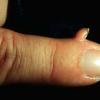
Clinically, acquired digital fibrokeratomas manifest as solitary, skin-colored, dome-shaped papules or tall fingerlike protrusions with a hyperkeratotic surface. Most acquired digital fibrokeratoma lesions are small and do not exceed 1.5 cm in height or diameter, but giant lesions measuring in excess of 3 cm have been documented.8
An important clinical finding reported to help differentiate acquired digital fibrokeratomas from other similar lesions is a collarette of slightly raised skin that encircles the base of the lesion, thereby creating a moatlike configuration
Causes
The etiology of acquired digital fibrokeratomas is unknown. Although trauma has been implicated, no studies can substantiate this hypothesis.
One report describes familial occurrence of an acral fibrokeratoma variant that had mucinous deposition; however, the case reported by Moulin et al10 histologically more closely resembles a superficial acral fibromyxoma.
Histologic Findings
The overall architecture of an acquired digital fibrokeratoma is a small, well-circumscribed, dome-shaped or narrow elongated papule. The stratum corneum is typically hyperkeratotic, which tends to be most pronounced toward the summit of the lesion. The epidermis can be acanthotic, with elongation of the rete ridges or can be slightly attenuated.
The dermal core of an acquired digital fibrokeratoma displays 1 of 3 histological patterns, which were originally described by Bart et al1 and further characterized by Kint et al.14 Type I acquired digital fibrokeratoma is the most common type and consists of a dermal core composed of thick, closely intertwined collagen bundles that are often oriented along the vertical axis of the lesion. Between the collagen bundles are numerous capillaries, varying numbers of fibroblasts, and thin elastic fibers. Type II lesions are less common, and histologically resemble type I lesions, but in addition have a significantly increased number of fibroblasts arranged in fascicles and markedly reduced numbers of elastic fibers. Type III acquired digital fibrokeratoma is the least common type and consists of a dermal core that is poorly cellular and edematous with a reduced number of elastic fibers
Surgical Care
Simple excision of acquired digital fibrokeratomas is curative; recurrence is rare.
.