| Atypical moles=شامات لا نموذجية |
|
|
Atypical moles In 1820, Norris proposed an association between nevi and melanoma. He described a family in which 2 members developed melanoma, while other family members had "many moles on various parts of their bodies." However, the exact appearance of these lesions is unknown. In 1974, Munro1 described an association of lesions and a family history of melanoma. These nevi had the clinical and microscopic appearance of what are now called atypical moles (AMs). In 1978 and 1981, Clark et al2,3 described these lesions as dysplastic nevi when they were observed as a familial phenomenon.
Atypical moles and dysplastic nevi are acquired melanocytic lesions of the skin whose clinical and histologic definitions are controversial and still evolving. Numerous definitions and criteria have been proposed, including the use of the term atypical moles for clinically abnormal nevi and dysplastic nevi for histologically abnormal nevi. Unfortunately, when clinically abnormal nevi are evaluated histologically, some studies have shown a lack of concordance, with some clinically abnormal nevi having no dysplastic features and some normal-appearing nevi having some dysplastic features. Atypical moles differ from common acquired melanocytic nevi in several respects, including diameter and lack of pigment uniformity. Confusion exists because some atypical moles cannot be clinically distinguished from melanoma. The clinical and histologic appearances of atypical moles occurring in a familial setting appear to overlap with sporadically occurring atypical moles. The US National Institutes of Health Consensus Conference on the diagnosis and treatment of early melanoma defined a syndrome of familial atypical mole and melanoma (FAMM). The criteria for FAMM syndrome are as follows9 :
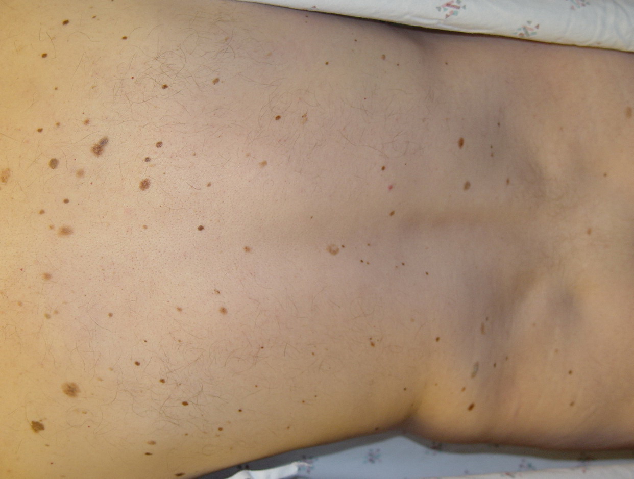
For additional information on malignant melanoma, see Malignant Melanoma. Additionally, the Medscape Skin Cancer Resource Center and Melanoma Resource Center may be helpful. PathophysiologyAtypical moles can be inherited or sporadic. Formal genetic analysis has suggested an autosomal dominant mode of inheritance but genetic studies have not shown consistent data. Germline mutations in 3 genes, CDK2NA10 and CDK4,11 mapped to 9p21 and 12q14, and CMM1, mapped to 1p,12 have been linked to a subset of hereditary melanomas and FAMM syndrome. In addition, somatic mutations in PTEN, BRAF,13 and MCR1 (melanocortin-1 receptor)14 have been associated with melanoma. Other genomic events such as loss of heterozygosity (LOH) for tumor suppressor genes are also responsible for the progression from atypical nevi to melanoma,15 and the genes thought to be responsible for most familial and sporadic atypical moles are still unknown. Ultraviolet (UV) light (UV-A and UV-B) has been proposed as both an initiator and a promoter in the transformation of melanocytes into atypical melanocytes or melanoma. The International Agency for Research on Cancer raised the classification of UV-emitting tanning devices from "probable carcinogenic to humans" to "carcinogenic to humans,"16 and a meta-analysis concluded that use of UV tanning beds before age 30 years increases the risk of melanoma by 75%.17 UV light exposure may be required for full expression of FAMM syndrome.Genetics and UV radiation may also result in a variable number and anatomical distribution of melanocytic nevi. Some patients with the atypical mole syndrome have many large and highly atypical nevi, whereas other patients with this syndrome have many nevi but only a few are atypical. FrequencyUnited StatesThe prevalence of atypical moles in white populations has been reported to be as high as 17%.18 Atypical moles can be inherited or occur sporadically. Familial atypical moles may be inherited as an autosomal dominant trait. Sporadic lesions are those atypical moles that occur in patients without a family history of atypical moles.19 InternationalIn Australia and New Zealand, the prevalence of atypical moles has been reported to be 5-10%.20 In Germany, approximately 2% of 500 white males aged 16-25 years were reported to have atypical moles on biopsy analysis. Eighteen percent of a population of white adults studied in Sweden were determined to have atypical moles clinically, although only 8% demonstrated histologic features of atypical moles. The marked differences in prevalence between different populations may be due to true differences between these populations or they may be related to differing clinical and histologic definitions of this entity. Mortality/MorbidityMelanoma can develop from precursor nevi and atypical moles. While many melanomas arise de novo, superficial spreading melanoma may arise from atypical moles.21 The exact risk of an individual nevus transforming into a melanoma is thought to be 1 in 200,000, and cutaneous melanomas are associated with precursor lesions at least 50% of the time in patients younger than 30 years.22 Patients with numerous atypical moles are at a higher risk of developing melanoma than those individuals with only a few atypical moles. This risk is more pronounced with a family history of melanoma.
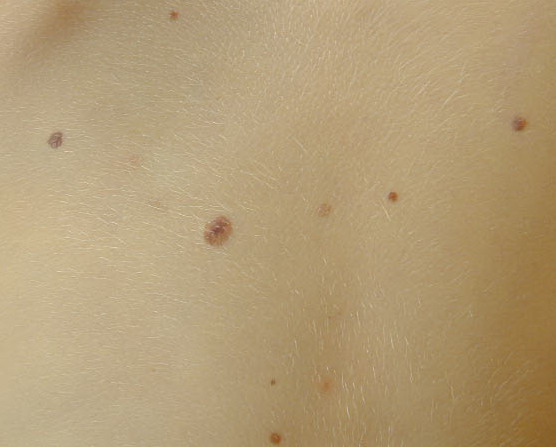
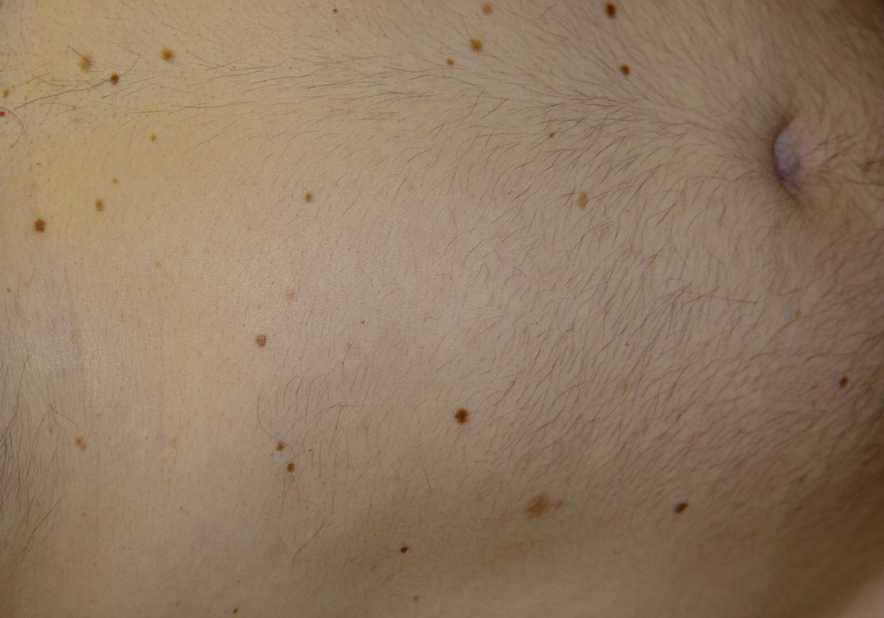
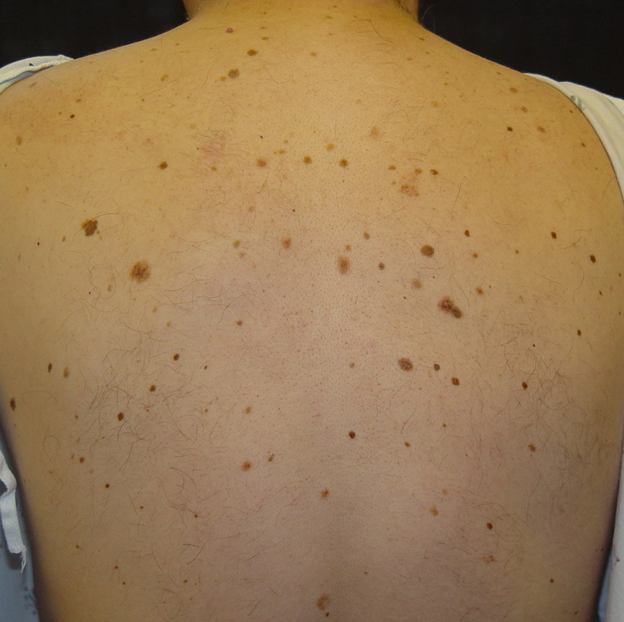
RaceIndividuals at the highest risk of atypical moles are persons of northern European background (Celtic) with light-colored hair and freckles. Atypical moles are rare in black, Asian, or Middle Eastern populations. SexNo sexual predilection is reported for atypical moles. AgeIn familial atypical moles, lesions begin to develop in childhood, most frequently during the first decade of life. Lesions may not be clinically specific early on, but typical features usually develop by the end of puberty. ClinicalHistoryA detailed personal and family history should be obtained, with special attention regarding moles and melanomas. PhysicalPatients with FAMM syndrome should have a complete cutaneous examination performed at the first office visit and then at least every 12 months for life. Atypical moles often have a characteristic appearance, although individual lesions may not show all the findings. Typically, they are large pigmented lesions and frequently measure 5-15 mm in diameter. Atypical moles are usually larger than common moles. Borders are usually irregular, notched, and ill defined. Macular and papular areas may be present within a single lesion (also described as a "fried egg" appearance). Color is highly variable and ranges from tan to dark brown to pink.18 CausesAtypical mole may be inherited (FAMM syndrome) or appear sporadically.27 Sun exposure may play a part in the distribution patterns of these nevi, but it is not absolutely necessary because atypical moles also appear on sun-protected skin. Patients with FAMM syndrome are at an increased risk for the development of melanoma, although the individual risk is variable
Histologic FindingsTypical histopathologic features, which are superimposed on those of a typical junctional or compound nevus, include the following (Note: Some clinical atypical moles are normal histologically):
The above changes may appear focally in any given lesion, and they may not be evident unless multiple histopathologic sections are studied. The World Health Organization Melanoma Program has proposed a similar list of characteristics/criteria for dysplastic nevi. Criteria are divided into 2 major and 4 minor criteria. An individual lesion requires 2 major and 2 minor criteria to be classified as a dysplastic nevus.5 Currently, most dermatopathologists are not using this classification scheme. However, the establishment of widely accepted criteria may eventually result in the uniform selection of patients for trials and population studies.
TreatmentMedical CareAll patients diagnosed with 1 or more atypical mole (AM) should undergo a complete cutaneous examination. Patients should be taught self-examination to detect changes in existing moles and to recognize clinical features of melanomas. Several studies have shown that regular cutaneous examinations combined with baseline and serial color photographs of the patient's cutaneous surface ultimately decrease biopsies and lead to earlier diagnoses of melanoma.26 Surgical CareBecause melanomas may develop de novo on the skin and because the risk of any one atypical mole developing malignant transformation is low, the prophylactic removal of all atypical moles does not prevent the development of melanoma and is not recommended. Changing lesions and any lesion worrisome for melanoma must be removed. . |