| Angioma serpiginosum= الوعاؤوم الساعي |
|
|
Angioma serpiginosum Angioma serpiginosum is an uncommon cutaneous vascular nevus of superficial capillaries characterized by minute puncta in clusters or in a linear array (a serpiginous pattern). These puncta result from a congenital hyperplasia or ectasia of preexisting superficial dermal capillaries, which may ultimately disappear (probably as a result of thrombosis). Electron microscopic findings have supported the view that these lesions are due to a vascular anomaly rather than a simple telangiectasia.
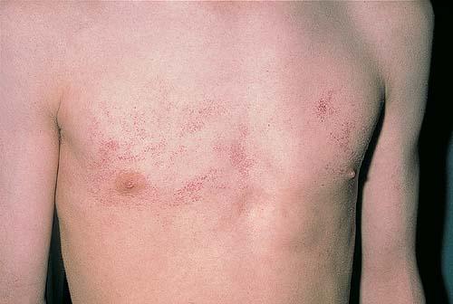
ClinicalHistoryAngioma serpiginosum, a rare vascular nevoid disorder due to ectatic dilation of capillaries in the papillary dermis, is found almost exclusively in females. In 2005, Sandhu and Gupta7 reported 2 rare cases—one with familial involvement and the other with an extensive distribution of lesions. Affected individuals tend to have grouped erythematous punctate lesions on the lower limbs or buttocks. Note the following:
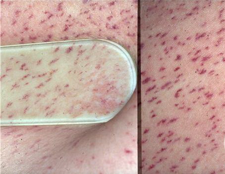
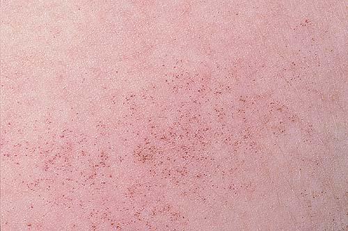
PhysicalAngioma serpiginosum is composed of reddish-purple puncta that may be as large as 1 mm. They are usually found grouped on the lower extremities in a serpiginous pattern. Rarely, the sole may be involved. Punctate erythematous maculae on the backs of the hands, arms, and shoulders may appear following a pregnancy. Angioma serpiginosum is variably compressible. The lack of inflammation, hemorrhage, or hemosiderin pigmentation is characteristic. Diascopic pressure applied to the lesion may produce only partial emptying, with some small tufts distended by purple venous blood remaining unchanged.
Histologic FindingsThe overlying epidermis is normal. The dermal papilla and subpapillary regions of the dermis show dilated capillaries with a thickening of the capillary walls. No inflammatory changes, hemorrhage, or hemosiderin depositions are present. Ultrastructural analysis of several lesions has shown that some thickening of vessel walls may occur from a heavy precipitate of basement membrane–like, fine fibrillar material mixed with thin collagen fibers. Some of these dilated capillaries show slitlike protrusions of lamina into the endothelial lining.
TreatmentMedical CareThis vascular lesion does not require medical care. Surgical CareElectrolysis or laser surgery of an individual lesion may be beneficial. Good cosmetic results can be achieved with a tunable pulse dye laser by selective photothermolysis of the vascular ectasias.12,18,19 With the tunable pulse dye laser, good-to-excellent results may be achieved in 4 or fewer visits |