|
Rheumatoid nodule = عقيدة رثوانية |
 |
 |
 |
|
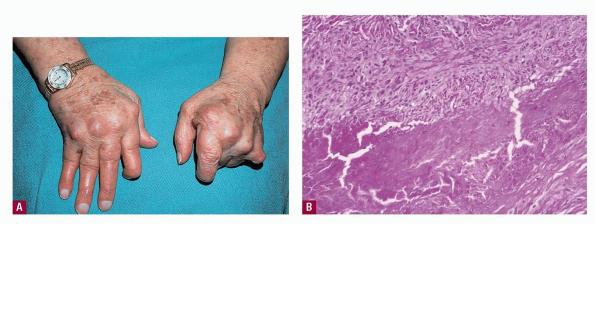
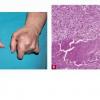
Rheumatoid nodules are deeply seated firm masses that occur in patients with rheumatoid arthritis, particularly over extensor prominences, such as the proximal ulna, the olecranon process, and the metacarpophalangeal and proximal interphalangeal joints . They may occur elsewhere, such as the back of the hands, over amputation stumps , and, rarely, in extracutaneous sites, such as the lung and heart . The nodules vary in size from a few millimeters to 5 em and may be solitary or numerous . Rarely, rheumatoid nodules show a central draining perforation . Rheumatoid factor is almost always found in high titer. Rarely, nodules may precede apparent joint disease . The rapid appearance of many small rheumatoid nodules has been reported in some patients treated with methotrexate. This presentation has been termed accelerated rheumatoid nodulosis
. The term rheumatoid nodulosis has been proposed for the clinical presentation of multiple nodules on the hands/elbows with intermittent arthralgias/arthritis but no evidence of systemic rheumatoid arthritis . Rheumatoid nodules also occur in occasional patients with systemic lupus erythematosus who do not exhibit rheumatoid arthritis .
|
|
Pseudorheumatoid nodule is a term that has been applied to nodules in the subcutis that mimic rheumatoid nodules histologically but that develop in the absence of joint pain, rheumatoid arthritis, or systemic lupus erythematosus . These occur in children or adults. The subsequent development of rheumatoid arthritis occurs infrequently in adults and rarely, if ever, in children. Because some of these nodules occur in patients with other lesions that are typical of intradermal granuloma annulare , the nodules are now generally considered to represent a subcutaneous variant of granuloma annul are.
|
|
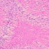
Histopathology.
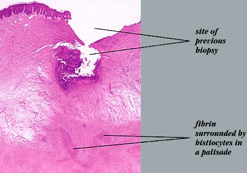
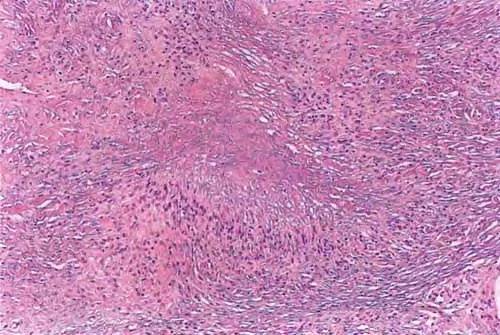
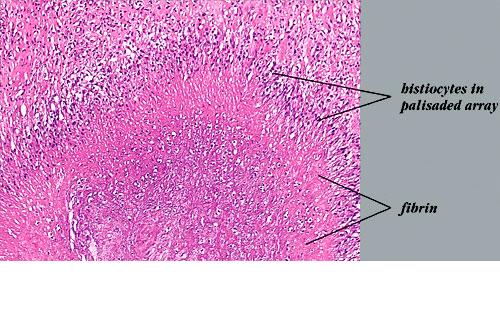
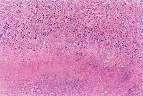
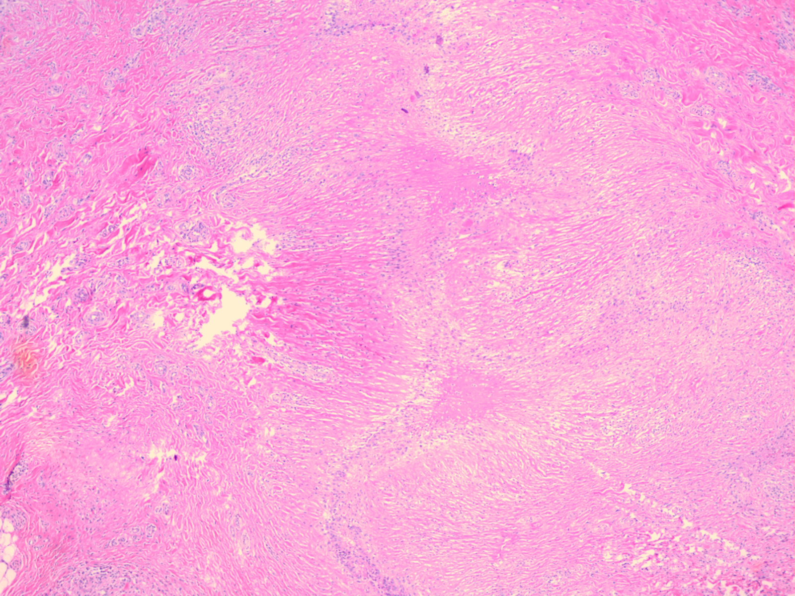
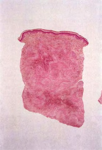
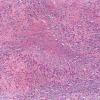
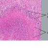
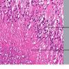
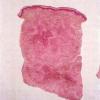
Rheumatoid nodules occur in the subcutis and deep dermis. They exhibit one or several areas of fibrinoid degeneration of collagen that stain homogeneously red . Nuclear fragments and basophilic material are often present, but mucin is almost always minimal or absent . These foci of degenerative change are surrounded by histiocytes in a palisade. Foreign-body giant cells are present in approximately 50% of biopsies . In the surrounding stroma, there is a proliferation of blood vessels associated with fibrosis. A sparse
infiltrate of other inflammatory cells is associated with the histiocytes and surrounding stroma. Lymphocytes and neutrophils are most common, but mast cells, plasma cells, and eosinophils may be present. Occasionally, lipid is seen . Vasculitis has been described but is
|
|
not usually encountered . In perforating rheumatoid nodules, the central fibrinoid material connects to the overlying skin surface .
|
|
Pathogenesis. Factors that have been implicated in the formation of rheumatoid nodules include trauma, vasculitis, and a specific T-cell-mediated immune reaction .
|
Differential Diagnosis. The principal differential diagnosis is subcutaneous granuloma annulare, which was discussed in the section on granuloma annulare. A distinction should be made from epithelioid sarcoma, which was also covered in that section. Nonabsorbable sutures or other foreign material may produce periarticular palisaded granulomas like those of rheumatoid nodule ; in such instances, there should be a history of previous surgery or trauma, and birefringent material may be visible under polarized light. Rheumatic fever produces nodules (rheumatic nodules), especially over the elbows, knees, scalp, knuckles, ankles, and spine , which were confused with rheumatoid nodules in the early part of the 20th century. Histologically, a rheumatic fever nodule is less likely to show central, homogeneous fibrinoid necrosis. A palisade of histiocytes is usually not as well developed, and fibrosis is minimal or absent . Rarely, an infectious process, such as cryptococcosis, can produce a deep, palisaded granuloma. It can be differentiated from rheumatoid nodule because the palisade surrounds primarily necrotic debris and organisms rather than fibrinoid material
|