|
Malignant blue nevus (melanoma) =الوحمة الزرقاء الخبيثة_ميلانوما |
 |
 |
 |
|
Malignant Blue Nevus
There exist potentially malignant melanocytic neoplasms that are unusual and do not necessarily qualify for application of the prognostic attributes of conventional vertical-growth-stage melanoma. These include the malignant blue nevus and a conceptual family of neoplasms of uncertain biological/malignant potential discussed in the next section. Malignant blue nevus is a rare tumor. It may arise in a blue or cellular blue nevus , a giant congenital nevus , or in a nevus of Ota , or it may be malignant from the start (765). Malignant blue nevi may involve the dermis and may be ulcerated or may present as a deep-seated expansile mass (765). In some lesions classified as malignant blue nevus, metastases occur that are limited to the regional lymph nodes, and the patient survives after removal of the tumor and the involved lymph nodes . There may be overlap in these cases with the phenomenon of cellular blue nevi with regional metastases. In other cases, however, death occurs as the result of widespread metastases . Unfortunately, reliable distinction between these two groups of cases is not possible.
|
|
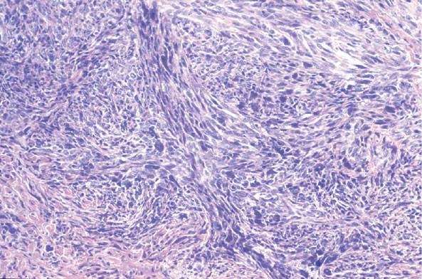
Histopathology.
Recognition of the lesion as a malignant blue nevus rather than a common melanoma is based on the absence of junctional activity and the presence of at least some bipolar tumor cells with branching dendritic processes typically containing melanin granules, representing a background component of blue nevus or cellular blue nevus . In some cases, pigment may be scant.
|
|
In addition to showing the standard features of malignancy, such as invasiveness of the tumor, atypia with hyperchromatism, pleomorphism and irregularity of the nuclei, and presence of atypical mitoses, malignant blue nevi often show areas of necrosis as evidence of their malignant nature . The combination of uniform cytologic atypia, high-grade atypia, spontaneous tumor necrosis, and more than a few mitoses may be considered diagnostic of malignant blue nevus in a lesion with a characteristic associated blue nevus pattern. Such cases are easily recognized as fully malignant neoplasms. Some lesions that do not meet all of these criteria; for example, some lesions that have lacked necrosis have metastasized . Atypical tumors with the overall appearance of cellular blue nevus that show only some of these features, especially if these are present in minor degree, may be signed out descriptively as melanocytic tumor of uncertain potential (see later section).
|
|
Histogenesis. Although some authors have regarded the tumor cells as related to Schwann cells , electron microscopic studies have shown the presence of melanosomes in the cells and a lack of cytoplasmic enclosures of unmyelinated axons, which would be seen if the tumor cells were Schwann cells. Although the melanosomes in many cells are devoid of melanin , incubation with DOPA has shown that they are strongly DOPA positive . Thus, it is evident that the tumor cells are melanocytes. Genetic data regarding the pathogenesis of malignant blue nevi are not available.
|
|
Differential Diagnosis. Malignant blue nevus differs from conventional primary melanoma by the absence of junctional activity and the presence of associated common and/or cellular blue nevus. However, distinction of a malignant blue nevus from a metastatic melanoma can be difficult because metastatic melanoma is occasionally found without a demonstrable primary melanoma. The primary melanoma may have involuted or may be located at an obscure internal site. The presence of dendritic cells indicative of an associated blue nevus or cellular blue nevus component is then the most reliable criterion favoring a diagnosis of malignant blue nevus instead of a metastatic melanoma. In doubtful cases, the differential diagnosis of metastatic melanoma should be expressed, and a workup for another primary should be considered clinically.
|
|