
Glomus Tumor
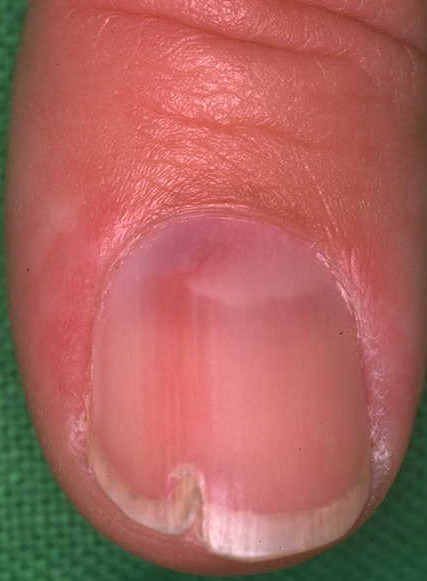
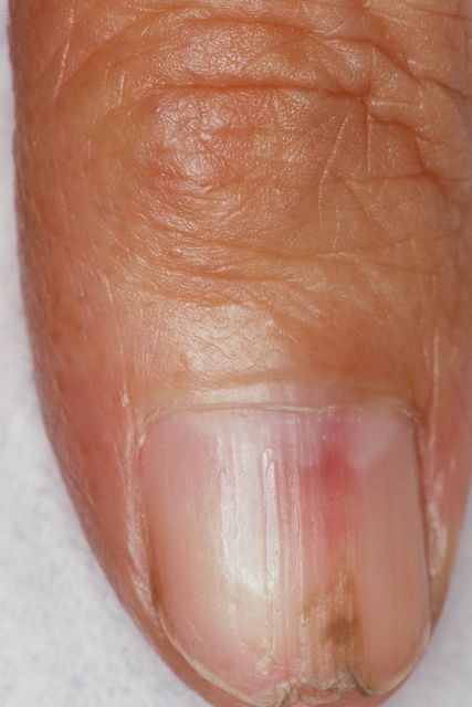
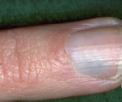
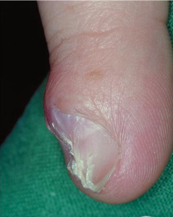
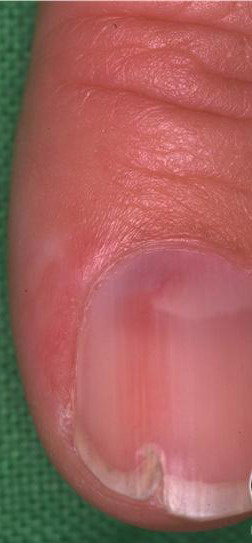
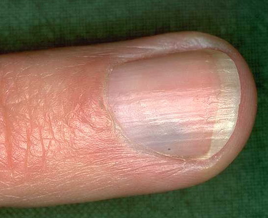
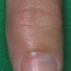
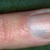
Glomus tumors are relatively uncommon benign neoplasms that differentiate to become modified smooth muscle cells called glomus cells. Two variants are noted: solitary glomus tumors and multiple glomus tumors, which are also known as glomangiomas or glomulovenous malformations. Each variant has distinct clinical and histopathologic characteristics. The most common location for these tumors is the distal extremities, especially in subungual areas.
Glomus tumors arise from the arterial portion of the glomus body, or the Sucquet-Hoyer canal, which is an arteriovenous shunt in the dermis that contributes to temperature regulation. Although glomus tumors are thought to arise from glomus cells, these tumors have been observed in extracutaneous locations that are not known to contain glomus cells. One explanation for this finding is that these tumors may arise from perivascular cells that can differentiate into glomus cells. Multiple glomus tumors, especially the disseminated variant, are inherited in an autosomal-dominant pattern with incomplete penetrance.
The exact incidence of glomus tumors is unknown. The multiple variant is rare, accounting for less than 10% of all cases. The probable misdiagnosis of many of these lesions as hemangiomas or venous malformations also makes an accurate assessment of incidence difficult.
The most common adverse effect is pain, which is usually associated with solitary lesions. Multiple tumors are less likely to be painful. In one report, a patient with more than 400 glomus tumors had thrombocytopenia as a result of platelet sequestration (ie, Kasabach-Merritt syndrome). Malignant glomus tumors, or glomangiosarcomas, are extremely rare and usually represent a locally infiltrative malignancy. However, metastases do occur and are usually fatal.
Solitary glomus tumors, particularly subungual lesions, are more common in females than in males. Multiple lesions are slightly more common in males.
Solitary glomus tumors are more frequent in adults than in others. Multiple glomus tumors develop 10-15 years earlier than single lesions; about one third of the cases of multiple tumors occur in those younger than 20 years. Congenital glomus tumors are rare; they are plaquelike in appearance and are considered a variant of multiple glomus tumors.
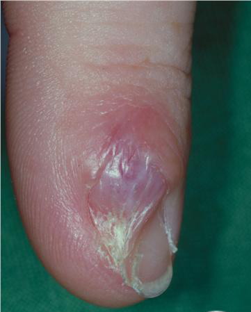
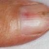
- Patients with solitary glomus tumors usually have paroxysmal pain, which can be severe and exacerbated by pressure or temperature changes, especially cold.
- Multiple glomus tumors can also be painful, but this feature is less common, and the pain is usually not severe.
- Patients with multiple lesions often seek medical attention because they are worried or have cosmetic concerns.
- Because multiple glomus tumors are inherited as an autosomal-dominant condition, a family history of similar lesions may be helpful for diagnosis.]
- Gastric glomus tumors are rare but have been reported.1
- Solitary glomus tumors have the following characteristics:
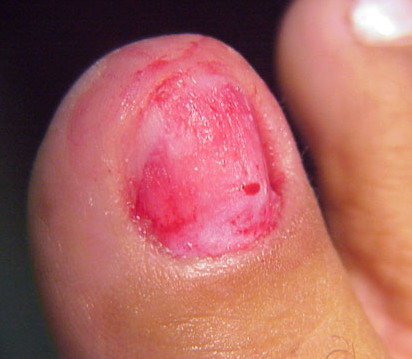
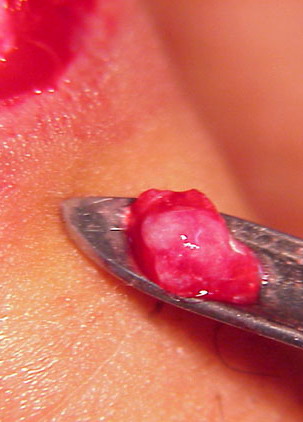
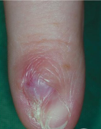
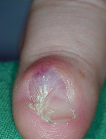
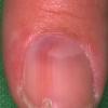
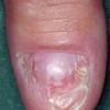
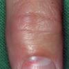
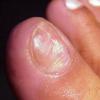
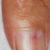
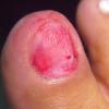
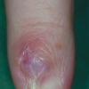
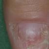
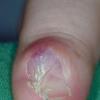
- Blue or purple
- Papules or nodules that can be blanched
- Size usually smaller than 1 cm
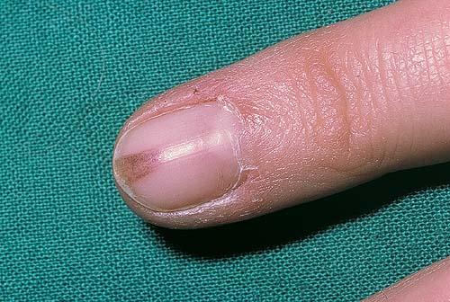
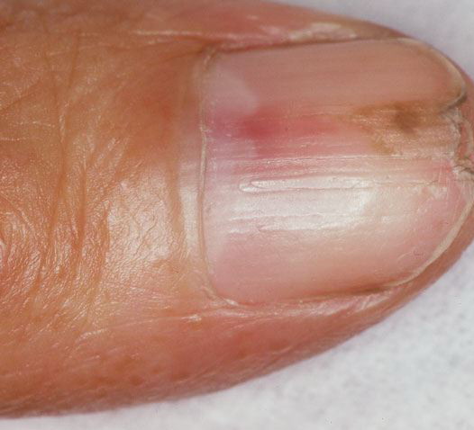
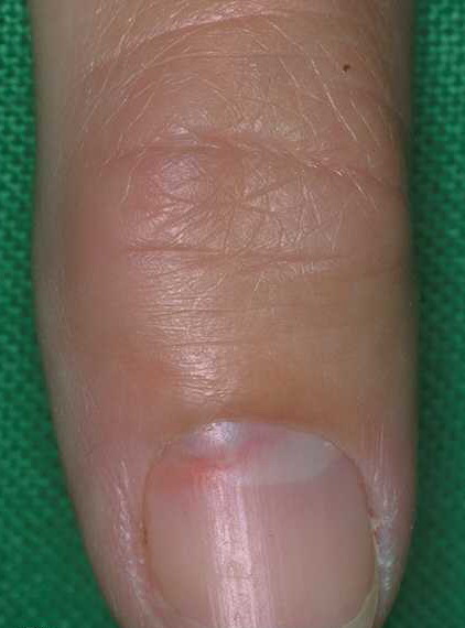
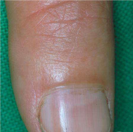
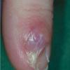
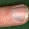
- Located most commonly in acral areas, especially subungual areas of fingers and toes
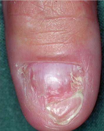
- The multiple variant is subdivided into regional or localized, disseminated, and congenital plaquelike forms.
- The regional variant consists of blue-to-purple partially compressible papules or nodules that are grouped and limited to a specific area, most commonly an extremity.
- The disseminated type consists of multiple lesions distributed over the body with no specific grouping. This form is less common than the regional variant.
- Congenital plaquelike glomus tumors consist of either grouped papules that coalesce into indurated plaques or clusters of discrete nodules. This form is the rarest variant of multiple glomus tumors.
- Two useful items for diagnosing glomus tumors, particularly solitary painful glomus tumors (especially those under a nail) are the following:
- Hildreth sign, which is disappearance of pain after application of a tourniquet proximally on the arm
- Love test, which consists of eliciting pain by applying pressure to a precise area with the tip of a pencil
- Features of glomangiosarcomas may include the following:
- Size larger than 1 cm
- Rapid growth
- Deep soft tissue involvement
- Glomus tumors are neoplasms caused by a proliferation of glomus cells, which make up a portion of the glomus body.
- The initiating event for glomus cell proliferation is unknown.
- Some authors have postulated that trauma induces solitary subungual glomus tumors, although this theory is not well studied.
- Most multiple glomus tumors, especially those of the disseminated form, are inherited in an autosomal dominant pattern with incomplete penetrance. Most hereditary glomangiomas are associated with defects in the glomulin gene, located on chromosome 1.
Treatment
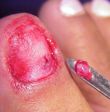
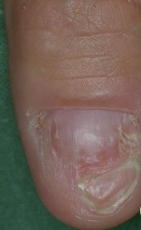
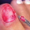
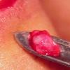
- The treatment of choice for solitary glomus tumors is surgical excision.
- For multiple glomus tumors, excision may be more difficult because of their poor circumscription and the large number of lesions.
- Excision should be limited to symptomatic lesions.
- Other reported treatment modalities include argon and carbon dioxide laser therapy and sclerotherapy with hypertonic saline or sodium tetradecyl sulfate. These are most useful in treating multiple lesions.
- The treatment of glomangiosarcoma is based on a few case reports.
- Wide local excision is adequate treatment and probably the treatment of choice.
- However, geometric excision is probably a reasonable alternative in cosmetically sensitive areas.
-