| Hidrotic ectodermal dysplasia = عسر تصنع الوريقة الظاهرة التعرقي |
|
|
HIDROTIC
ECTODERMAL
DYSPLASIA
Epidemiology
Hidrotic ED was first described in a French-Canadian kindred. It has been reported in other ethnic groups, but the majority of affected individuals can trace their ancestry back to an original French-Canadian settler. Box 143-1 Differential Diagnosis of Some Diagnostic Features of Ectodermal Dysplasias
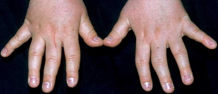
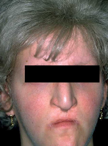
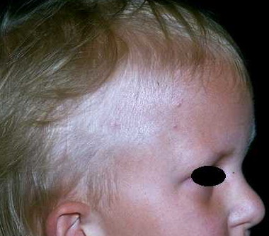
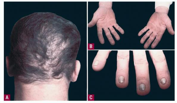
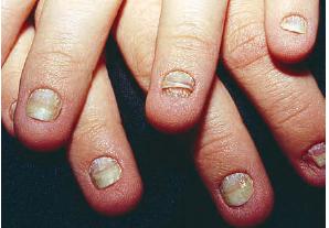
Etiology, Pathogenesis, and Genetics The disorder is caused by mutations in a connexin gene, GJB6 or connexin-30.17 Different mutations in the same gene are responsible for a form of nonsyndromic autosomal dominant deafness and at least one patient with keratitis-ichthyosis-deafness (KID syndrome) (see Chap. 47). Other connexin genes show similar variability in mutation: disease correlations [e.g., mutations in connexin-31 (GJB3)] can cause either erythrokeratodermia variabilis (see Chap. 47) or late-onset autosomal deafness. The pathway by which allelic mutations result in such different diseases is not yet known. Hidrotic ED is autosomal dominant with variable expression (the degree of severity can vary within and between families). Males and females are affected in equal numbers and to equal degree. The gene maps to the centromeric region of the long arm of chromosome 13. Clinical Manifestations The scalp hair is wiry, brittle, and pale, and there is often patchy alopecia . This progresses in adult life and may lead to total alopecia. Body and facial hair are affected. The nails may be milky white in infancy and early childhood, gradually thickening and becoming dystrophic. The nail plates in adults are thick, short, slow-growing, separate distally from the nail bed , and may cause pain. Anonychia has been reported. Not all the nails are necessarily affected to the same degree. Progressive palmar/plantar hyperkeratosis is common . In contrast to HED, sweating is normal, as are the teeth. Oral leukoplakia has been reported. Conjunctivitis and blepharitis, possibly due to poor function of sparse eyelashes, are common.
Histopathology The thickened palms and soles show orthohyperkeratosis with a normal granular layer. On electron microscopy, an increase in the number of desmosomes in the cells of the stratum corneum is found. The hair shows non-specific changes. Diagnosis and Differential Diagnosis The diagnosis is straightforward. The involvement of nails and hair and palmar/plantar thickening, in the absence of other signs of ED, are reasonably specific. Other palmar/plantar hyperkeratoses do not have similar hair changes. Orofacial clefting differentiates other forms of autosomal dominant hidrotic ED, such as ankyloblepharon filiforme adnatum (AFA)-ED-cleft palate (AEC) syndrome or Rapp-Hodgkin syndrome. Although the nail changes are similar to those of pachyonychia congenita, the hair changes are distinctive. Treatment Occasionally, ablation of the nail matrix is necessary for relief of pain. Wigs may provide cosmetic benefit. Treatment of the thickened palms and soles is not specific and minimally successful.
|