|
Dermal naevu s= وحمة أدمية |
 |
 |
 |
 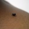 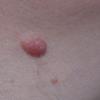 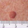
Dermal naevus
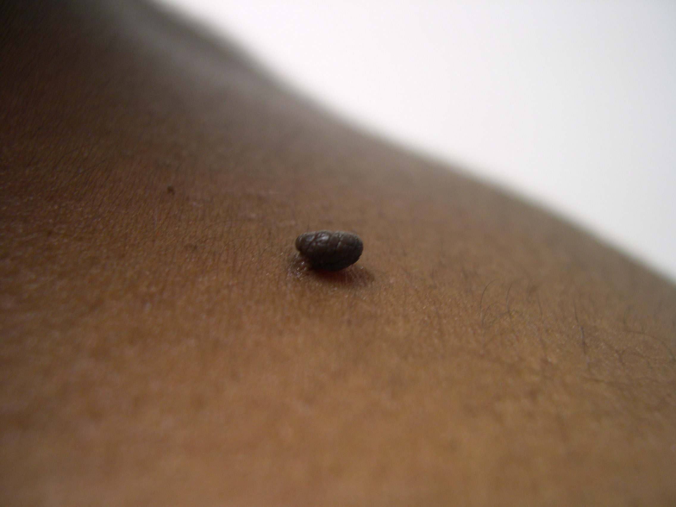
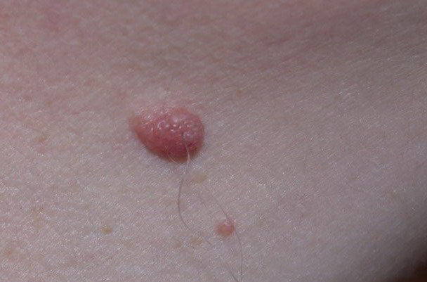
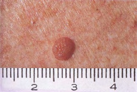
The DERMAL NEVUS represents the most mature stage of a MELANOCYTIC NEVUS. Its MELANOCYTES have almost totally been replaced by NEVOCYTES, "neuroid" cells that have formed cells clusters within deeper layers of the skin. They are MOLES AT REST, and usually don't change their aspects over time - they are the final versions of a NEVUS. Consequently, they have lost their potential to malignify.
DERMAL NEVI are most frequently located on the facial skin, the scalp region, and the trunc. They are always palpable, usually less intensely pigmented than COMPOUND NEVI, and may exhibit hair growth within and keratotic plaques on the surface. Despite the fact that they usually don't look attractive and might thus be bothersome to many individuals, they aren't risky at all. Excsional biopsy and histological clarification is rarely required.
|