| Chondrodermatitis nodularis helicis=التهاب الجلد والغضروف العقيدي في الاذن- |
|
|
▪ CHONDRODERMATITIS
NODULARIS HELICIS
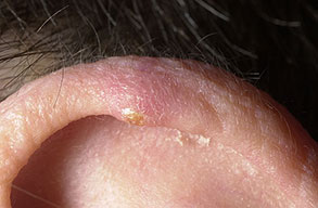
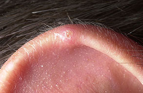
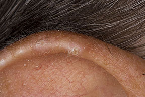
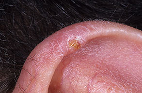
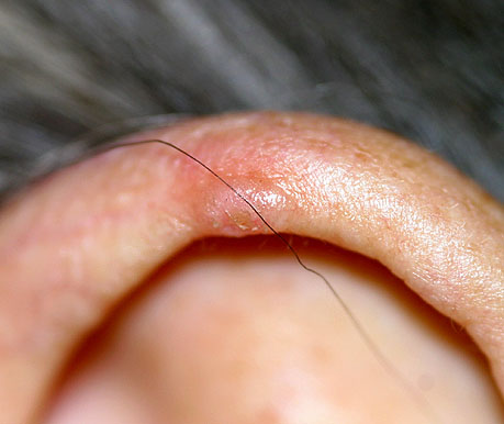
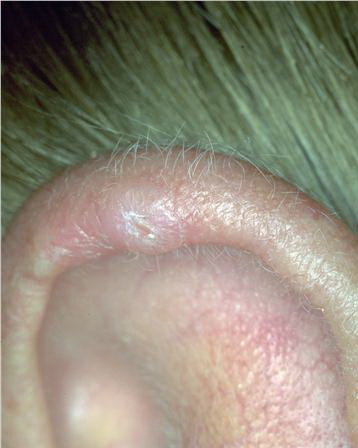
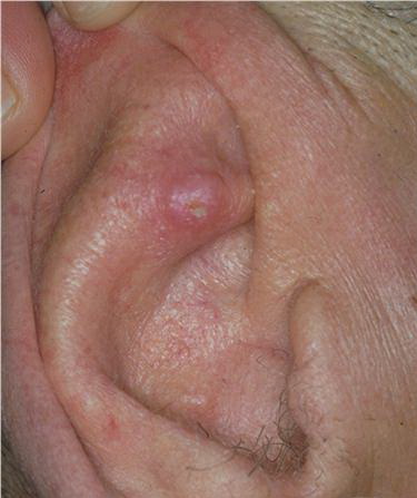
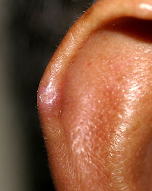
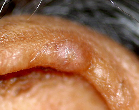
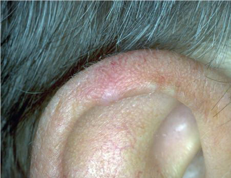
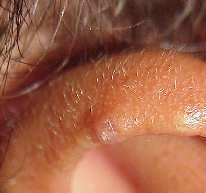
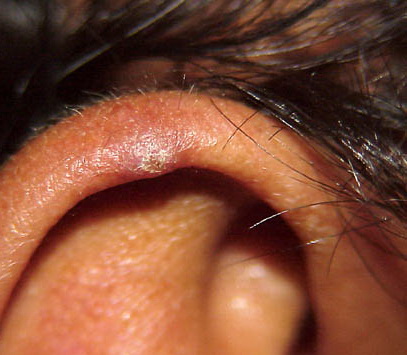
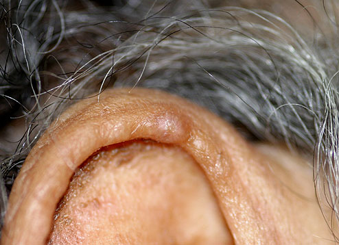
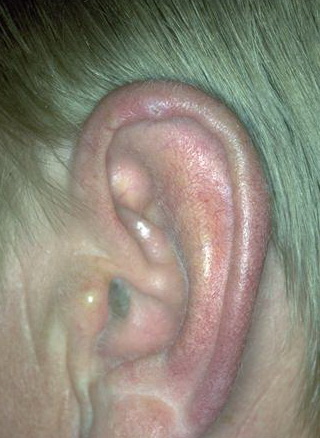
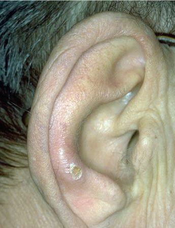
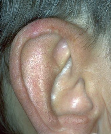
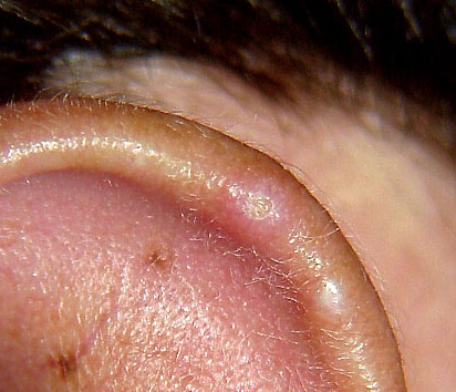
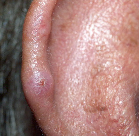
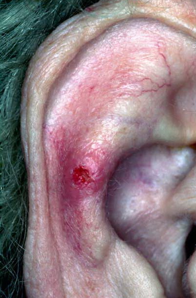
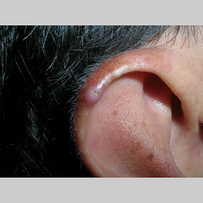
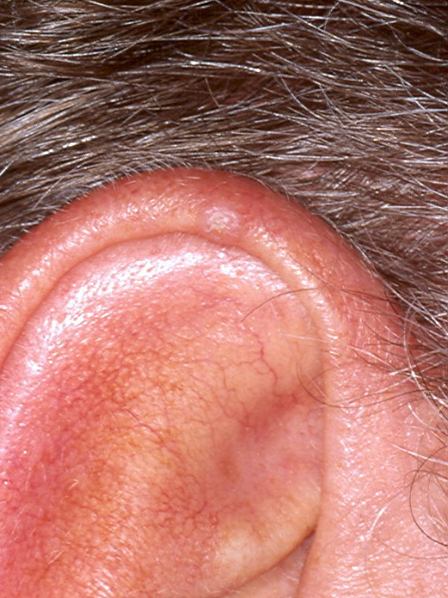
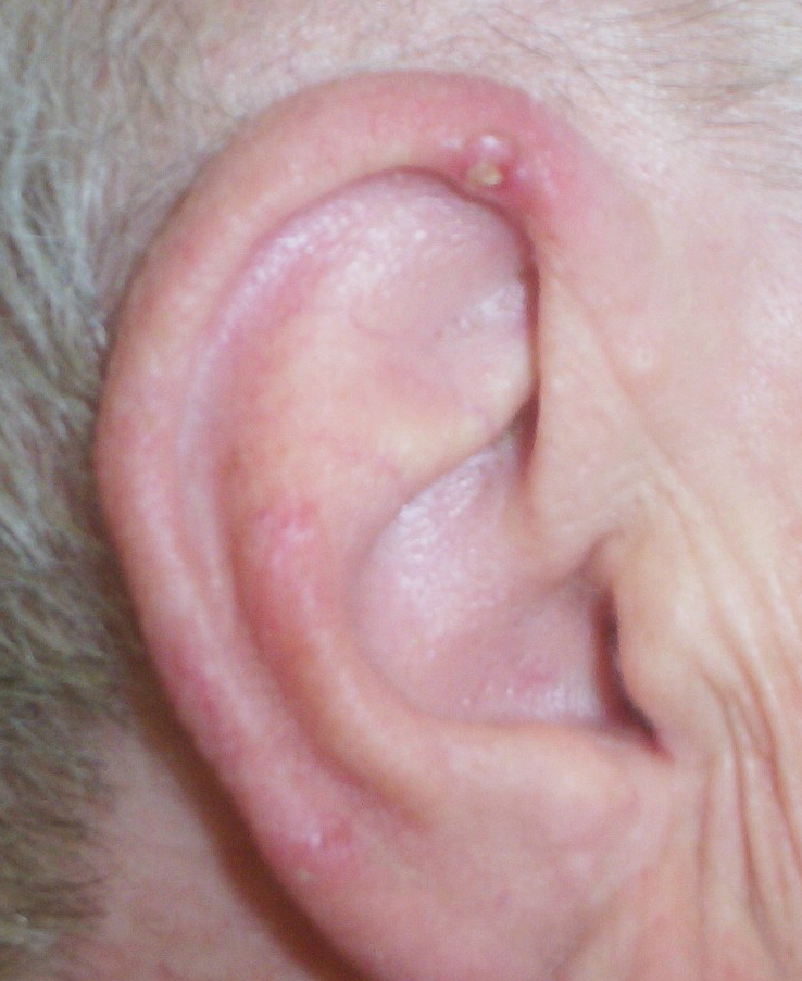
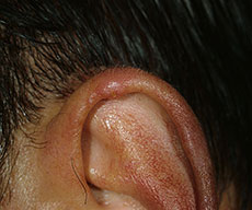
Clavus helicis CNH is a chronic, solitary, tender, ulcerated nodule on the helix of the ear, first described in 1915. Etiology and Pathogenesis Many etiologic factors have been suggested for CNH. It is thought to be a disease of degenerated collagen that undergoes trans-epithelial elimination.125 Vascular insufficiency, pressure, injury to the cartilage, sun-exposure, and low temperature have all been implicated in inciting cartilage damage. Recently, Magro et al. described 24 patients with early-onset CNH who had concomitant microvascular disease, strengthening the argument for the role of ischemia in the development of this process.126 Reports
CHONDRODERMATITIS NODULARIS HELICIS AT A GLANCE
CLINICAL FEATURES
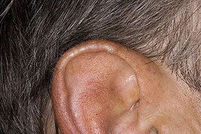
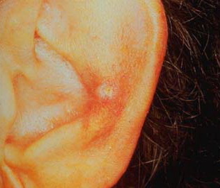
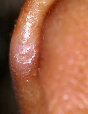
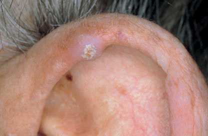
This disorder is found overwhelmingly in adult men over the age of 50, with the male-female prevalence being greater than 2:1. Two cases of CNH in children have been reported. Patients present with a history of a suddenly appearing, skin-colored to pink papule or nodule on the ear that grew rapidly and subsequently remained quiescent. These lesions tend to be on the right helix, measure less than 1 cm, and are associated with pain . The pain can be paroxysmal or associated with changes in ambient temperature. The lesion may have a central ulceration with an adherent hemorrhagic or scale crust. Spontaneous resolution of CNH is unlikely. A possible subset of CNH may be seen in elderly women who have a history of prior trauma. For these women, spontaneous regression is not uncommon.
Differential Diagnosis
Basal cell carcinoma, keratoacanthoma, warts, rheumatoid nodules, squamous cell carcinoma, clavus, and tophus are all within the differential of CNH. Pathology In the epidermis, there is commonly hyperkeratosis and parakeratosis; ulceration may be present. Occasionally, degenerated collagen is seen in the epidermis, indicating a trans-epidermal elimination process. A brisk, predominantly lymphocytic infiltrate is noted in the dermis with admixed histiocytes and few neutrophils. Telangiectasias and solar elastosis are often noted in the adjacent dermis. The perichondrium shows inflammation and fibrous thickening. The cartilage may show hyalinization, necrosis, and, at times, ossification.
Treatment
Conservative treatment includes pressure-relieving pillows with a “doughnut” shape that are commercially available. In a recent study examining the role of such pressure-relieving devices, 13 of 15 patients (87 percent) were healed at follow-up after 1 month of conservative treatment. High-potency steroids, intralesional steroids, and laser ablation may also be used to induce symptom control or regression of the lesion.134 Excision of CNH nodules using a shave or excisional technique through and around the affected cartilage is also an accepted treatment option, though recurrences may occur at the edge of the lesion.
|