| Carcinoma erysipelatoides= كارسينوما حمرانية |
|
|
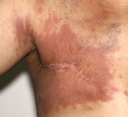
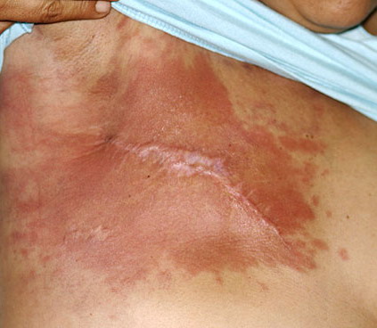
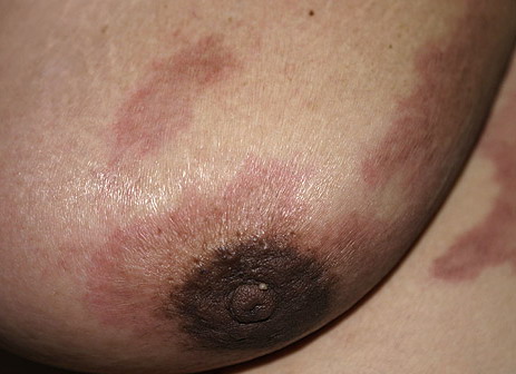
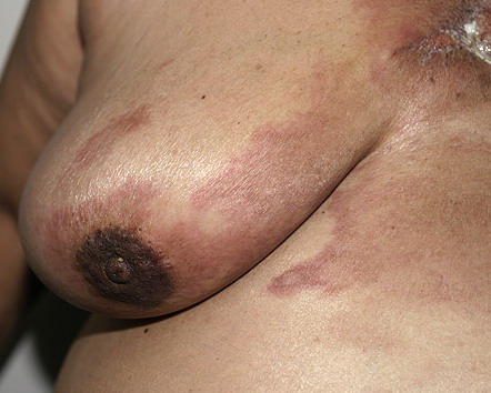
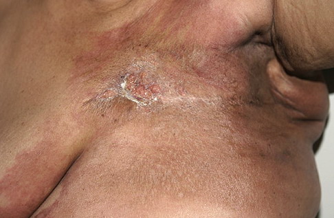
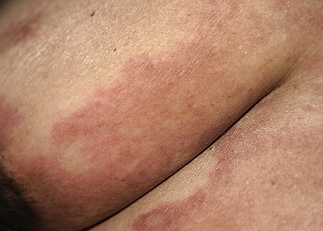
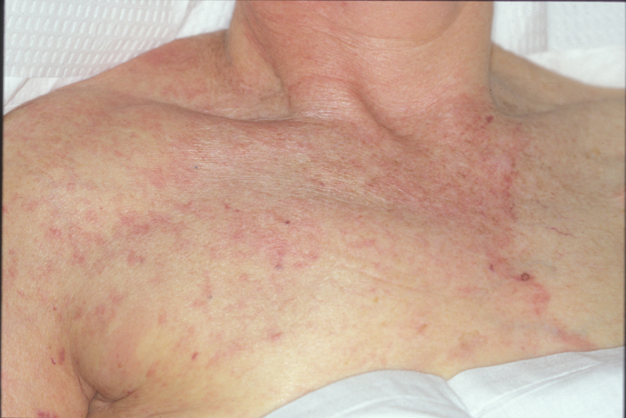
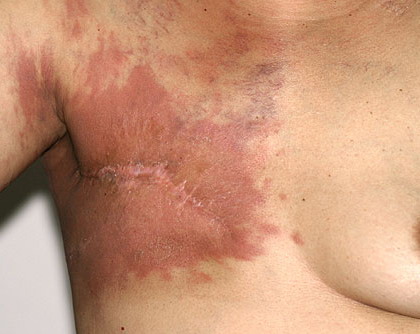
Carcinoma erysipeloides Carcinoma erysipeloides (CE), which clinically resembles erysipelas, is an uncommon, but distinctive form of cutaneous metastasis. It has been termed inflammatory metastatic carcinoma. It classically presents as a rapidly evolving unilateral chest wall erythema, which may extend to the back, proximal part of the arm and even across the midline
Histopathologic and immunohistochemical findingsIn all skin biopsy specimens, histopathologic features of adenocarcinoma with varying degrees of cell differentiation were observed. The most frequent histopathologic findings were single or multiple nodules located in the dermis and subcutaneous tissue, com posed of small to large aggregates of tumor cells surrounded by fibrosis. Neoplastic cells were typically arranged in gland-like structures (Fig. 4) or in a linear distribution between collagen bundles in an "indian file" pattern. Telangiectatic carcinoma was characterized by aggregates of tumor cells and erythrocytes as well as dilated blood vessels in the papillary dermis. In erysipeloid carcinoma, histopathologic features were metastatic cells tightly packed within dilated superficial and deep lymphatic vessels and a slight perivascular infiltrate of lymphocytes and plasma cells. In carcinoma "en cuirasse", fibrosis with few neoplastic cells sometimes exhibiting a characteristic "indian file" pattern was detected. Histopathologic findings of nodular carcinoma or "en cuirasse" carcinoma associated with atrophy of the hair follicle as a result of fibrosis were observed in metastatic lesions of the scalp. Finally, an epidermotropic pattern characterized by neoplastic cells, single or in small nests, within the epidermis and dermal aggregates of neoplastic cells in a sclerotic stroma was identified in 7/164 patients (4%).Immunohistochemical investigations showed in all patients a strong positivity of tumor cells for pan-cytokeratins (PKK1, AE1/AE3, LU5, CK) and epithelial membrane antigen (EMA) (Fig. 5). In addition, a positive reaction for carcinoembryonic antigen (CEA) was detected in 110/164 of the cases (67%).
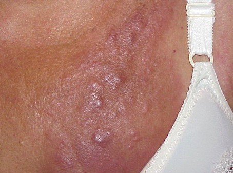
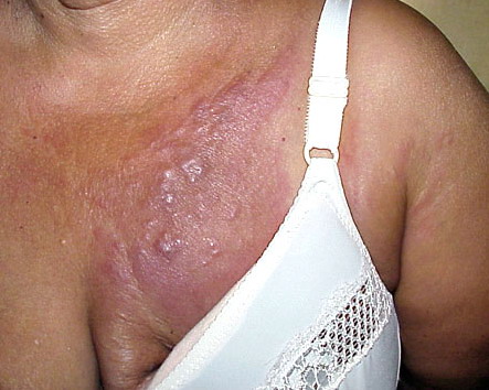
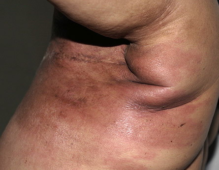
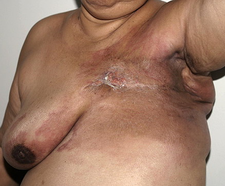
DiscussionCutaneous involvement from breast carcinoma preferentially occurs in the skin overlying or proximal to the area of the primary tumor by direct extension or through lymphatic vessels. Nodular carcinoma, inflammatory or erysipeloides carcinoma, telangiectatic and "en cuirasse" carcinoma are the typical clinical manifestations of the lymphatic dissemination to the skin. Inflammatory carcinoma occurs when neoplastic cells disseminate through the lymphatics of the entire thickness of the dermis and subcutaneous tissue. In contrast, telangiectatic carcinoma is characterized by dissemination through superficial lymphatics and blood vessels of the dermis. In nodular carcinoma and "en cuirasse" carcinoma, the tumor cells disseminate largely along tissue spaces and only to a minor degree through lymphatic vessels (6). Although the above mentioned clinical manifestations are highly suggestive of cutaneous metastatic breast carcinoma, they may mimic a variety of benign and malignant cutaneous disorders as well as occur with cancers of other organs. Inflammatory skin metastases may clinically resemble erysipelas but, in contrast to true infection, there is no fever, chills and leukocytosis, and bacterial cultures are negative (7-11). This clinical pattern may also develop with cancers from other sites such as pancreas, colon and rectum, lung, ovary, prostate and parotid gland. Telangiectatic lesions have also been reported in carcinoma of the uterine cervix and parotid gland, and "en cuirasse" metastatic carcinoma may rarely be seen in kidney, lung and gastrointestinal malignancies. Hematogenous spread of neoplastic cells is responsible for alopecia neoplastica, which may simulate alopecia areata, sebaceous cyst, scleroderma, morphea-like basal cell carcinoma and discoid lupus erythematosusUnusual and nonspecific clinical appearances of cutaneous metastatic breast carcinoma have been described. A zosteriform eruption, as observed in one of our patients, has been rarely reported and most likely results from a perineural lymphatic metastatic dissemination (14,15). The dermatomal distribution of vesicular lesions may clinically resemble herpes zoster. However, the site of cutaneous lesions, the presence of malignant cells within the vesicles and a negative viral PCR or culture allow to establish the definite diagnosis of cutaneous metastasis. Metastatic breast carcinoma presenting as a reddish nodule on the tip of the nose has been described and defined "clown nose" for its peculiar clinical feature (16). In addition, breast cancer of the inframammary crease may appear, mainly in women with pendulous breast, as cutaneous exophytic nodules clinically suggestive of a primary cutaneous squamous or basal cell carcinoma (17). Finally, metastatic breast cancer in the eyelid has been reported as a painless swelling of the eyelid associated with induration or nodule formation. Histologically, it may show characteristics of ductal or pleomorphic carcinoma as well as a histioid appearance (18). Immunohistochemical studies may be helpful to identify the site of the primary tumor. In our series and previous reports, cutaneous metastatic breast carcinoma stained positively with antibodies to keratin proteins, as well as with anti-CEA and anti-EMA antibodies (6,19). In addition to estrogen and progesterone receptors, the gross cystic disease fluid protein-15 (GCDFP-15), a monoclonal glycoprotein expressed in apocrine epithelial cells and metaplastic apocrine tissue, has been described as a useful diagnostic marker for identifying primary as well as metastatic breast carcinoma (20-22). Recently, Bayer-Garners and Smoller suggested that androgen receptors might serve as additional immunohistochemical markers to increase sensitivity for detecting breast cancer in skin metastasis (23). The prognosis of patients with cutaneous metastasis depends on the type and biological behavior of the underlying primary tumor and on its response to treatment. Skin metastases from breast carcinoma are usually associated with advanced stages of the disease as observed in our study and, therefore, in most cases, they represent a poor prognostic sign. Systemic chemotherapy is the most commonly used treatment whereas the specific protocol depends on the histopathologic type of the primary tumor and staging of the patient. Surgical excision, radiotherapy, intralesional chemotherapy or immunotherapy can be used when solitary lesions develop or in the late stages of the disease in order to improve the quality of life of the patient. |