| Calcifications = التكلسات |
|
|
▪ CALCIFICATION
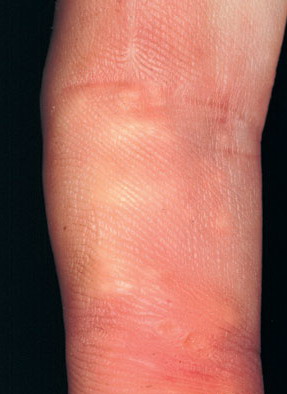
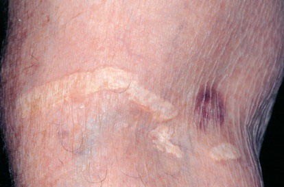
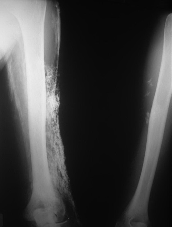
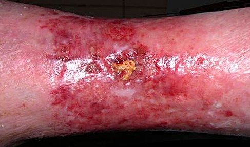
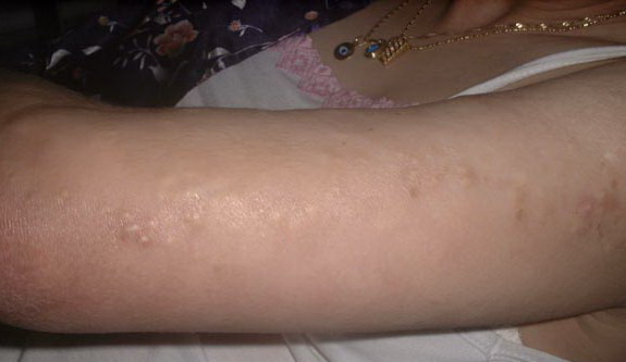
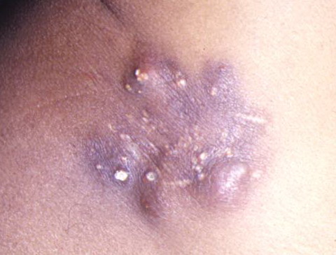
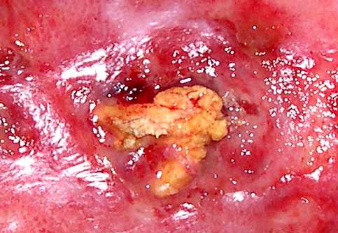
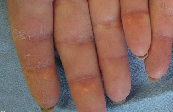
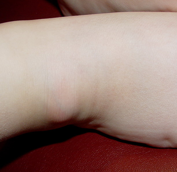
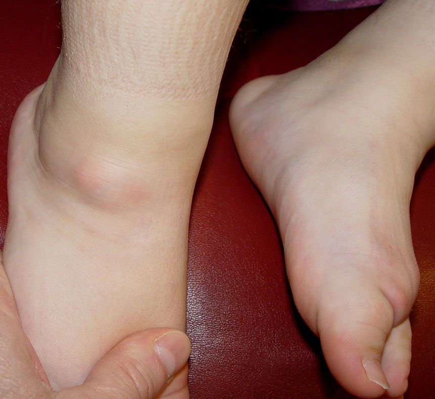
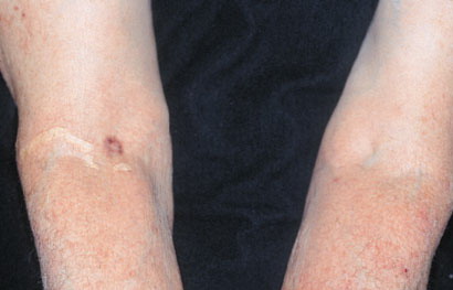
Connective Tissue Diseases Dystrophic calcification frequently occurs in connective tissue diseases. Scleroderma and CREST syndrome (calcinosis cutis, Raynaud phenomenon, esophageal dysfunction, sclerodactyly, telangiectasia) are notable examples that are frequently associated with calcinosis cutis . In these disorders, nodules and plaques of calcium deposits may occur in the skin, subcutaneous tissue, muscle, or tendons. The calcium deposits most commonly occur on the upper extremities, especially on the fingers and wrists, but may occur in any area subject to trauma or motion. As the calcifications enlarge, they may ulcerate and exude a chalky material. Dystrophic calcification also occurs in dermatomyositis . It is more commonly associated with juvenile rather than adult-onset dermatomyositis, occurring in 44 percent to 70 percent of children as opposed to 20 percent of adults. The calcification tends to occur 2 to 3 years after disease onset and most frequently appears on the elbows, knees, shoulders, and buttocks.18 The calcium deposits may be painful and can ulcerate. They also may exude a chalky material, form sinuses, and become chronically infected. Calcium salt deposition may become quite extensive, progressing along fascial planes of skin and muscle, forming an “exoskeleton,” and leading to significant morbidity and mortality. Calcinosis cutis in dermatomyositis is difficult to treat; however, if the patient survives long enough, the calcified nodules may improve spontaneously. Although uncommon, calcinosis cutis has been described in all clinical subsets of lupus erythematosus. There is no standard treatment for dystrophic calcification. A diet low in calcium and phosphate along with aluminum hydroxide has been reported to arrest or facilitate regression of the calcified nodules.23 Disodium etidronate has also been used with some success.24 Several series report long-term treatment with diltiazem improves the calcinosis in some patients.25,26 Other reported treatments include warfarin, colchicine, probenecid, and bisphosponates.16,27 Occasionally, calcium deposits must be removed surgically to clear sinus tracts, ulcers, or chronic infections. Panniculitis Pancreatic enzyme panniculitis is a lobular panniculitis that commonly demonstrates dystrophic calcification. It occurs in patients with pancreatitis or pancreatic adenocarcinoma and is presumably caused by the action of liberated pancreatic enzymes on subcutaneous fat. The fatty acids formed by lipolysis may combine with calcium and form calcium soap. In subcutaneous fat necrosis of the newborn, erythematous, well-defined nodules and plaques occur during the first few weeks of life over the cheeks, back, buttocks, and extremities. The affected infants are generally otherwise healthy, and the nodules and plaques usually clear spontaneously. Occasionally, the lesions calcify, and in a small subset of patients symptomatic hypercalcemia may develop, sometimes several months after birth.
Inherited Disorders
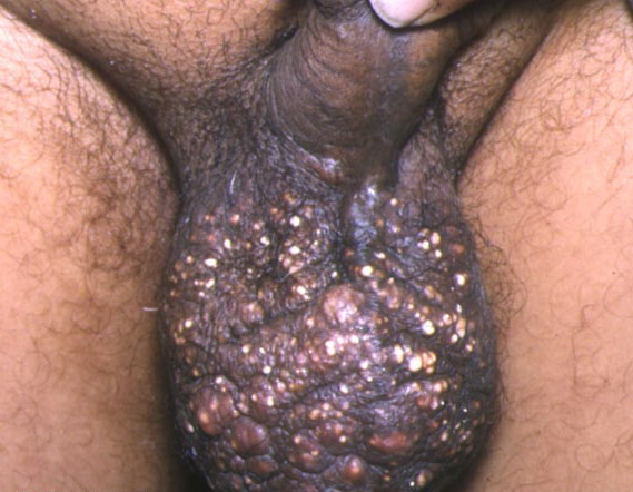
Dystrophic calcification occurs in patients with pseudoxanthoma elasticum . PXE is a hereditary disorder of elastic tissue characterized by progressive calcification of elastin fibers, primarily within the skin, Bruch's membrane of the retina, and the cardiovascular system. The cause of PXE was recently identified as a mutation in the ABC-C6 gene. This gene is thought to play a critical role in transmembrane transport. Most patients with PXE have normal calcium-phosphate metabolism, but a few have been identified who have abnormal calcium, phosphate, and/or vitamin D metabolism. Patients in this subset may develop metastatic calcification in the form of calcified or ossified tumors, calcification of the falx cerebri, and arterial calcification.31-33 Ehlers-Danlos syndrome is a group of inherited disorders of fibrillar collagen metabolism. Mutations in the collagen genes or enzymes that regulate collagen biosynthesis have been determined to underlie a number of EDS sub-types.34-36 The skin characteristically shows hyperelasticity and fragility with formation of pseudotumors and large gaping scars. Subcutaneous calcified nodules, termed spheroids may appear, and are thought to represent calcified ischemic fat lobules.37,38 Calcification of healing surgical incisions has also been reported in patients with EDS. Dystrophic calcification has been observed in patients with porphyria cutanea tarda Sclerodermoid plaques with dystrophic calcification have occurred on the preauricular area, scalp, neck, and dorsa of the hands. Ulceration with trans-epidermal elimination of sheets of calcium is also rarely reported. Other genetic disorders in which calcification may occur include Werner syndrome and Rothmund-Thomson syndrome
Cutaneous Neoplasms
Dystrophic calcification occurs in association with a variety of benign and malignant cutaneous neoplasms. Often the neoplasms also show ossification in the surrounding stroma. Pilomatricomas are the most common cutaneous neoplasms that manifest calcification and ossification. Approximately 75 percent of pilomatricomas show calcification and 15 percent to 20 percent show ossification. Ossification usually occurs within the connective tissue adjacent to the shadow cells, probably through metaplasia of fibroblasts into osteoblasts. Activating mutations in the adherens junction protein β-catenin have been identified in some pilomatricomas.46 A large number of other neoplasms may be associated with calcification and ossification, including: pilar cyst, basal cell carcinoma, intradermal nevi (probably as a result of inflammation or folliculitis), desmoplastic malignant melanoma, atypical fibroxanthoma, pyogenic granuloma, hemangioma, neurilemmoma, trichoepithelioma, and seborrheic keratoses. Mixed tumors (chondroid syringomas) may also show calcification and ossification. However, unlike other neoplasms, the ossification occurs within the tumor via ossification of the chondroid cells, much like endochondral bone formation occurring in the epiphyses of bones.
Infections
Infectious agents may produce enough cutaneous damage to cause dystrophic calcification. Parasitic infections that may result in calcinosis cutis include onchocerciasis (Onchocerca volvulus) and cysticercosis (Taenia solium).52,53 Calcinosis cutis has also been reported as a complication of intrauterine herpes simplex infection.54 Other Dystrophic calcification has been reported in a variety of settings where local tissue injury occurs, such as in scarring caused by burns, trauma, neonatal heel sticks (sub-epidermal calcified nodule), surgery, and keloids.
|