Blepharochalasis
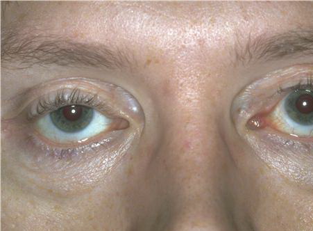
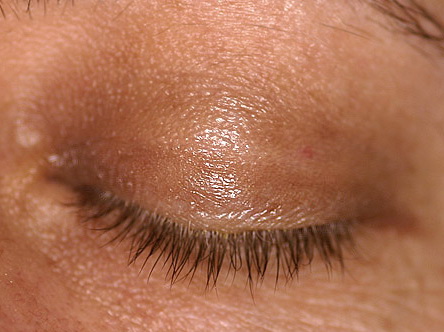
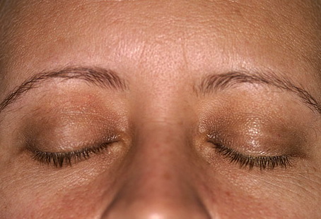
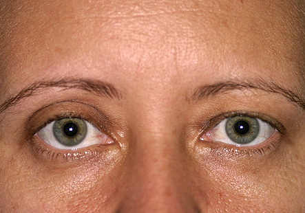
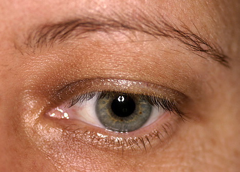
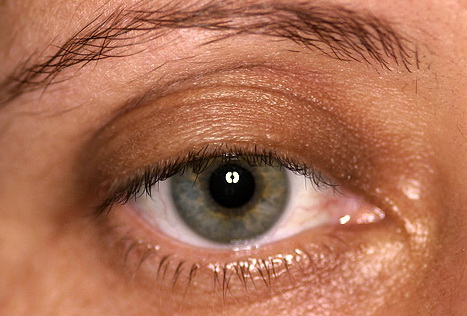
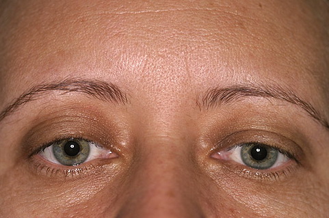
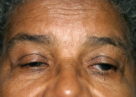
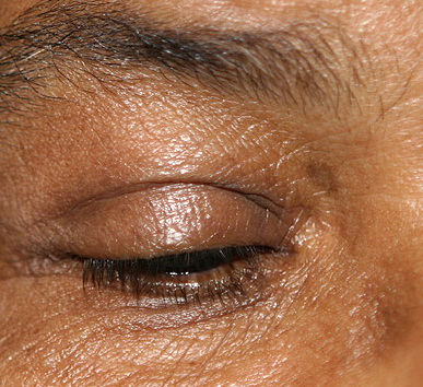
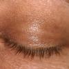
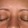
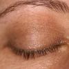
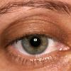
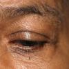
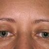
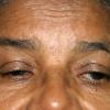
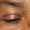
Blepharochalasis is a rare syndrome consisting of recurrent bouts of upper eyelid edema associated with thinning, stretching, and fine wrinkling of the involved skin. The lower eyelids are not commonly involved. These episodes often result in eyelid skin redundancy. In 1817, Beer initially described the condition; however, in 1896, Fuchs first assigned the term blepharochalasis to this entity.1 The word blepharochalasis originates from the Greek blepharon (eyelid) and chalasis (a relaxing).
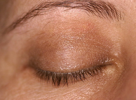
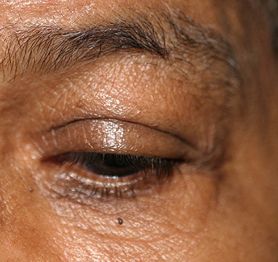
Various disease stages have been observed. In 1926, Benedict described a swelling stage and a subsequent stage characterized by thinning skin.5 Others have suggested an active, intumescent phase that precedes a quiescent, atrophic phase.
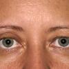
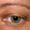
Blepharochalasis is often confused with dermatochalasis, which refers to the lax and redundant skin most commonly observed in the upper eyelids with aging. However, dermatochalasis is usually not associated with recurrent attacks of edema, “cigarette-paper” skin, and subcutaneous telangiectasia, as observed in blepharochalasis.
Blepharochalasis may be a form of chronic angioedema with localized vascular dilation and proteinaceous fluid extravasation. An orbital component has been suggested because, in patients with the syndrome, orbital fat has been noted to contain increased vascularity with dilated capillaries. Multiple triggers have been described, including immune reactions and environmental factors.7
The finding of immunoglobulin A (IgA) deposits in lesional skin has implicated immunopathogenic causes. Elevated immunoglobulin E (IgE) levels in one case report supports the involvement of atopy in blepharochalasis. Perivascular infiltrates in patients with active disease, along with degradation of both elastin and collagen in the dermis, suggest inflammatory influences. Elastin messenger RNA (mRNA) expression has been shown to be normal compared to controls, indicating an environmental cause of breakdown, such as postinflammatory enzymatic action.