|
Solar Elastosis
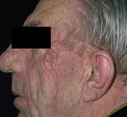
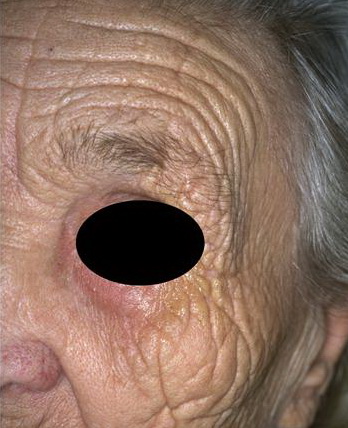
Senescent changes in areas of the skin not regularly exposed to sunlight manifest themselves clinically only in thinning of the skin and a decrease in the amount of subcutaneous fat. In contrast, there are often pronounced changes in the appearance of the exposed skin of elderly persons, especially those with fair complexions. These changes, however, are the result of chronic sun exposure (photoaging) rather than of intrinsic (chronologie) aging. In exposed areas, especially on the face, the skin shows wrinkling, furrowing, and thinning. In addition, there may be an irregular distribution of pigment.
|
|
Histopathology.
In skin not regularly exposed to sunlight, there is a progressive loss of elastic tissue in the papillary dermis with age. Elastic fibers in the papillary dermis are composed of elaunin and oxytalin fibers. Elaunin fibers, which consist of microfibrils and a small amount of elastin, run parallel to the dermal-epidermal junction. Overlying oxytalan fibers, composed solely of microfibrils, form a thin, superficial network perpendicular to the dermal-epidermal junction that ends at the basement membrane zone . In middle age, the oxytalan fibers in the papillary dermis are split and fewer than at a young age; in old age, they may be absent (1). Mature elastic fibers in the reticular dermis also undergo changes due to intrinsic aging, becoming fragmented and porous (2).
|
|
In the skin of the face exposed to the sun, especially in persons with fair complexions, hyperplasia of the elastic tissue is usually evident on histologic examination by the age of 30 years, even though clinically the skin may appear normal. No white person past 40 years of age has normal elastic tissue in the skin of the face (3). The elastic fibers of the reticular dermis are increased in number, and they are thicker, curled, and tangled.
|
|
In patients with clinically evident solar elastosis of the exposed skin, staining with hematoxylin-eosin reveals, in the upper dermis, basophilic degeneration of the collagen separated from a somewhat atrophic epidermis by a narrow band of normal collagen. In the areas of basophilic degeneration, the bundles of eosinophilic collagen have been replaced by amorphous basophilic granular material.
|
|
With elastic tissue stains, the areas of basophilic degeneration stain like elastic tissue and therefore are referred to as elastotic material. The elastotic material usually consists of aggregates of thick, interwoven bands in the upper dermis (4) but in areas of severe solar degeneration, the elastotic material may have an amorphous rather than a fibrous appearance and may extend into the lower portions of the dermis rather than being confined to the upper dermis (5).
|
|
On staining with silver nitrate, the distribution of melanin in the basal cell layer may appear irregular in that areas of hyperpigmentation alternate with areas of hypopigmentation (5).
|
|
Histogenesis. Electron microscopic examination of areas of solar elastosis shows elastotic material as the main component (EM 13). Even though this elastotic material resembles elastic tissue in its chemical composition, it differs significantly in appearance from aged elastic fibers in unexposed, aged skin. Instead of showing amorphous electron-lucent elastin and aggregates of electron-dense microfibrils (see Chapter 3), the thick fibers of elastotic material show two structural components: a fine granular matrix of medium electron density and, within this matrix, homogeneous, electron-dense, irregularly shaped inclusions (6). Microfibrils such as those observed in normal or aged elastic fibers are absent. Immunoelectron microscopy shows that the elastotic material has retained its antigenicity for elastin but not for microfibrils (7). The number and size of elastotic fibers are greatly increased over the number and size of elastic fibers found in normal or aged skin. Extensive amorphous material
|
|
can be seen around the elastotic fibers and also among the collagen fibrils. Collagen fibrils are diminished in number, with those present often showing a diminished electron density, a diminished contrast in cross striation, and a splitting up into filaments at their ends (8).
|
|
Elastotic material is not regarded as a degeneration product of preexisting elastic fibers. Most current findings indicate that elastotic material is composed primarily of elastic tissue, much of which may be newly formed as the result of an altered function of fibroblasts. Evidence of transcriptional activation of the elastin gene in biopsied tissue and fibroblast cultures from sun-damaged skin further supports this. Additional accumulation of elastotic material may be secondary to a disruption of the balance between synthesis and degradation of elastin in photodamaged skin (9).
|
|
The elastotic material that histochemically stains like elastic tissue resembles elastic tissue in its chemical composition and its physical and enzymatic reactions. Thus, the amino acid composition of the elastotic tissue resembles that of elastin and differs significantly from that of collagen. In particular, the
|
|
elastotic material, like elastic tissue, has a much lower content of hydroxyproline than collagen (10). Moreover, the elastotic material in unfixed sections shows the same brilliant autofluorescence as do elastic fibers on examination with the fluorescence microscope (11), and both the elastotic material and elastic tissue are susceptible to elastase digestion (12). The elastotic material contains a large amount of acid mucopolysaccharides, as indicated by staining with Alcian blue. A significant portion of these acid mucopolysaccharides may be sulfated because prior incubation with hyaluronidase removes only 50% to 75% of the Alcian blue-positive staining. The basophilia of the elastotic material, however, is not affected by incubation with hyaluronidase (13).
|
|
The irregular distribution of melanin in the epidermis observed in some patients with solar degeneration, when studied by electron microscopy, is found to be caused largely by an impairment of pigment transfer from melanocytes to keratinocytes. Although some keratinocytes contain many melanosomes, others contain few or no melanosomes. The latter are surrounded by dendrites laden with melanosomes (14).
|
|
Differential Diagnosis. For a discussion of differentiation of solar elastosis from pseudoxanthoma elasticum (PXE), see Chapter 3 and 6.
|
|
Localized Expressions of Solar Elastosis
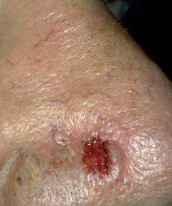
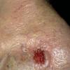
Several clinically distinct forms of localized solar elastosis have been described. In the nuchal region, the skin, after many years of exposure to the sun, may appear thickened and furrowed. This is referred to as cutis rhomboidalis nuchae. Elastotic nodules of the ears are localized papular and nodular forms of solar elastosis that usually occur on the antihelix (15, 16, 17,18). Severe solar elastosis may also occur as yellowish plaques associated with small cysts and comedones. Favre-Racouchot syndrome (nodular elastosis with cysts and comedones) is an example occurring on facial skin lateral to the eyes (19,20). A similar condition occurring on the arms has been termed actinic comedonal plaques (21 ,22,23). Two other types of circumscribed solar elastosis occurring on the upper extremities are solar elastotic bands of the forearm (4,24) and collagenous and elastotic marginal plaques of the hands (25,26,27,28,29,30,31 ,32).
|
|
Elastotic Nodules of the Ears
|
|
Elastotic nodules are most often seen on the anterior crus of the antihelix (15,16, 18) and occasionally on the helix of the ears (17). They are often bilateral. Clinically, they may mimic basal cell carcinomas, amyloidosis, gouty tophi, or chondrodermatitis nodularis helicis .
Histopathology. Irregular elastotic fibers and clumps of elastotic material are seen in the background of marked dermal solar elastosis (Fig. 15-2A). The fibers and clumps can be highlighted with a Verhoeff-van Gieson elastic stain (17) (Fig. 15-2B).
|
|
Favre-Racouchot Syndrome (Nodular Elastosis with Cysts and Comedones)
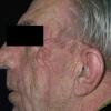
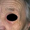
Favre-Racouchot syndrome is characterized by yellow plaques with multiple open and cystically dilated comedones. The condition typically affects the skin lateral to the eyes in elderly males (19,20,41). However, a case has also been documented on the shoulder (33). Although the condition is usually bilateral, it may be unilateral (34,35,36). It is thought to be primarily secondary to prolonged solar exposure with the formation of comedones facilitated by an extracellular matrix of compromised structural integrity (2). Smoking may also be a contributing factor in its development .
|
|
Histopathology. Dilated pilosebaceous openings and large, round, cystlike spaces are lined by a flattened epithelium and
|
|
represent greatly distended hair follicles (. Both the dilated pilosebaceous openings and the cystlike spaces are filled with layered horny material. Vellus hair shafts and bacteria have been demonstrated within the spaces as well, suggesting that the cystlike spaces may represent closed comedones rather than true infundibular cysts . The sebaceous glands are atrophic. Solar elastosis often is pronounced , but it may be slight or absent . Because the comedones are open, they do not tend to become inflamed .
|
|
Actinic Comedonal Plaques
In actinic comedonal plaques, solitary nodular plaques with a cribriform appearance and comedone-like structures occur, often unilaterally, on the arms or forearms . The plaques are composed of confluent erythematous to bluish papules and nodules. The condition has been described in fair-skinned individuals with a history of chronic sun exposure. They can be found in association with Favre-Racouchot syndrome and may in fact represent an ectopic expression of this entity .
Histopathology. Dilated corneocyte-filled follicular lumina are present within areas of elastotic, amorphous material. The overlying epidermis is usually dyskeratotic and atrophic. The histologic findings are quite similar to those seen in Favre-Racouchot syndrome .
|
|
Solar Elastotic Bands of the Forearm
|
|
Solar elastotic bands of the forearm consist of soft cordlike plaques across the flexor surface of the forearms . The bands occur in areas of actinic damage and usually with senile purpura.
|
|
Histopathology. Nodular collections of basophilic homogenous amorphous material underlying an atrophic epidermis are conspicuous features. Thickened degenerated elastic fibers within the homogenous material are also observed. Stellate fibroblasts and a perivascular infiltrate of lymphocytes and hemosiderin-laden macrophages are found in close apposition to the elastic fibers. The nodular collections and thickened elastic fibers stain positively with Verhoeff-van Gieson elastic stain (4).
|
|
Collagenous and Elastotic Marginal Plaques of the Hands
Collagenous and elastotic marginal plaques of the hands have been described by several names: elastocollagenous plaques of the hands , degenerative collagenous plaques of the hands (26,28), and keratoelastoidosis marginalis (27). This acquired, slowly progressive condition is usually seen in elderly males and consists of groups of linear confluent papules along the medial and lateral aspects of the hands at the juncture of the palmar and dorsal surfaces. The medial aspect of the thumb and radial aspect of the index finger are most commonly affected. The condition closely resembles the genodermatosis, acrokeratoelastoidosis . However, there is no familial predisposition or involvement of the plantar surfaces. Actinic damage and chronic repetitive pressure or trauma has been implicated in its pathogenesis .
Histopathology. The reticular dermis displays an acellular zone of haphazardly arranged collagen with some bundles running perpendicular to the epidermis . The bundles of collagen are admixed with fragmented elastic fibers and distinctive angulated amorphous "basophilic elastotic masses" in the upper dermis. These masses can be demonstrated to contain degenerating elastic fibers and calcium .
|
|