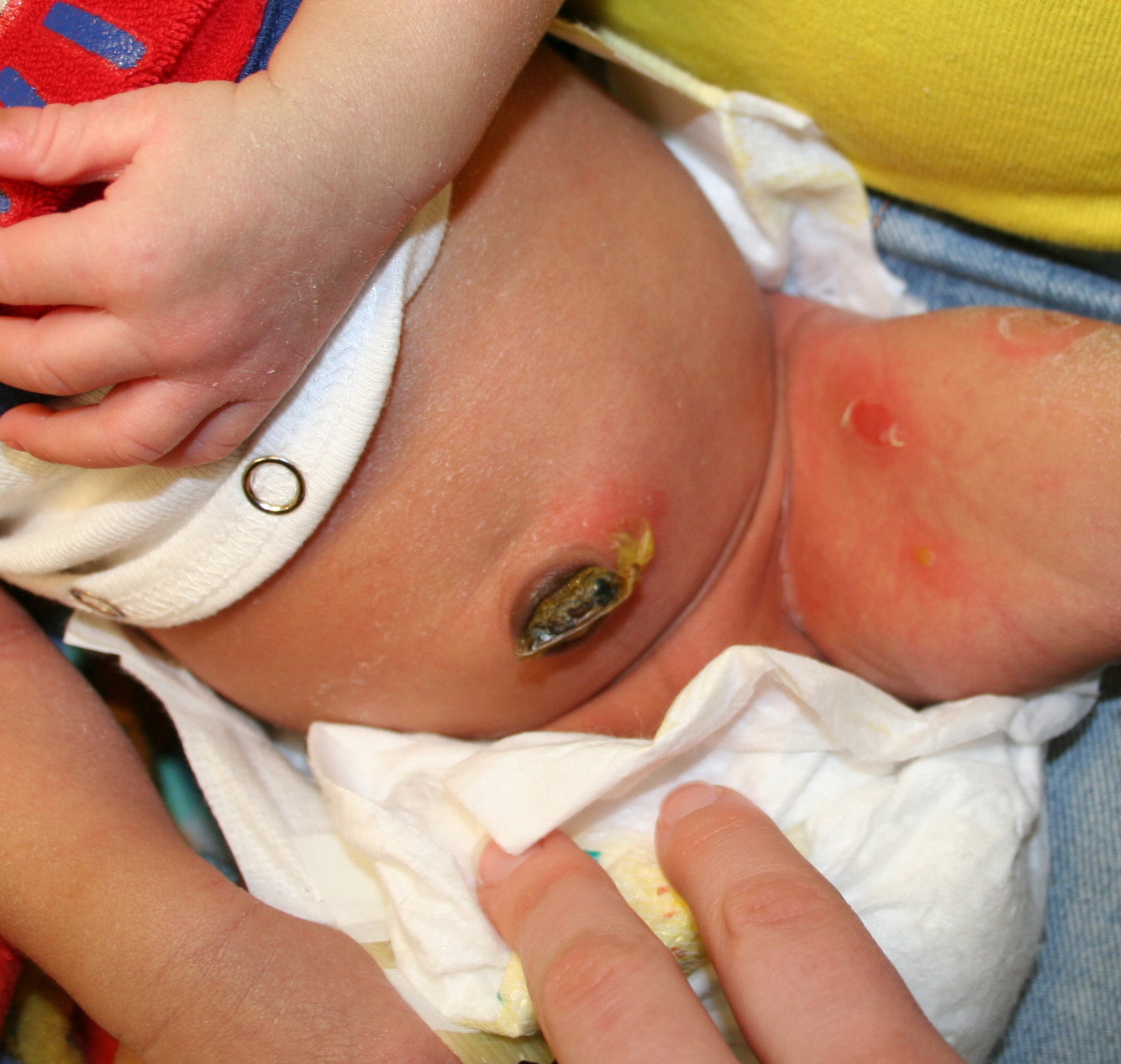
| Neonatal bullous impetigo = القوباء الفقاعية عند الوليد |
|
|
Impetigo.
Two clinical patterns of impetigo are recognized: bullous and non-bullous. Bullous impetigo is caused by S. aureus. Currently, in industrialized nations, non-bullous impetigo is most commonly caused by S. aureus and less often by group A streptococcus. Group A streptococcus remains a common cause of non-bullous impetigo in developing nations.
NON-BULLOUS IMPETIGO.
The non-bullous type of impetigo accounts for more than 70 percent of cases of this form of pyoderma. It occurs in children of all ages as well as in adults. Intact skin is usually resistant to colonization or impetiginization, possibly due to absence of fibronectin receptors for teichoic acid moieties on S. aureus and group A streptococcus. Production of bacteriocins, produced by certain S. aureus strains (phage group 71) and highly bactericidal to group A streptococcus, may be responsible for the isolation of only S. aureus from some lesions initially caused by streptococci.
History.
In a typical sequence, S. aureus spreads from nose to normal skin (approximately 11 days later) and then develop into skin lesions (after another 11 days). Lesions commonly arise on the skin of the face (especially around the nares) or extremities after trauma. Nasal carriers of S. aureus can present with a very localized type of impetigo confined to the anterior nares and the adjacent lip area ; pruritus or soreness of the area is a common complaint . Conditions that disrupt the integrity of the epidermis, providing a portal of entry of impetiginization, include insect bites, epidermal dermatophytoses, herpes simplex, varicella, abrasions, lacerations, and thermal burns.
Cutaneous Lesions.
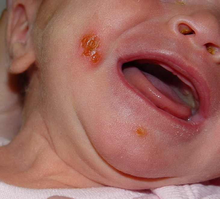
The initial lesion is a transient vesicle or pustule that quickly evolves into a honey-colored crusted plaque that can enlarge to greater than 2 cm in diameter . Surrounding erythema may be present. Constitutional symptoms are absent. Regional lymphadenopathy may be present in up to 90 percent of patients with prolonged, untreated infection. Untreated, the lesions may slowly enlarge and involve new sites over several weeks. In some individuals, lesions resolve spontaneously. In others, the lesions extend into the dermis, forming an ulcer (see Staphylococcal Ecthyma
|
||||||||||||||||||||||||||||||||||||||||||||||||||||||||||||||||||||||||||||||||||||||||||||||||||||||||||||||||||||||||||||||||||||||||||