| Arterial ulcer=قرحة شريانية المنشأ |
|
|
Arterial ulcer Ulceration due to vascular causes is often multifactorial and can be caused by both arterial and venous disease. Hypertension and atherosclerosis of the peripheral vessels lead to arterial disease associated with ischemic ulcers. Chronic venous insufficiency and the resulting venous hypertension cause venous ulcers. Vasculitis such as Buerger disease (thromboangiitis obliterans) or Takayasu disease can also be associated with ulceration. The former tends to manifest with arterial or ischemic-type ulcers, while the latter manifests with cutaneous disease such as pyoderma gangrenosum or erythema nodosum. When an ulcer does not respond to adequate medical and wound care, the potential for an underlying malignancy should be considered. Cutaneous malignancies that may masquerade as ulcers include nodulo-ulcerative basal cell carcinoma, squamous cell carcinoma, keratoacanthoma, nodular melanoma, tumor stage mycosis fungoides, lymphomatoid granulomatosis, lymphomatoid papulosis, angiosarcoma, and cutaneous metastases from internal malignancy. Healthcare providers must recognize these presentations and render appropriate therapeutic intervention.2 PathophysiologyArterial (or ischemic) ulceration can be caused by either progressive atherosclerosis or arterial embolization. Both lead to ischemia of the skin and ulceration. Venous (or stasis) ulceration is initiated by venous hypertension that develops because of inadequate calf muscle pump action and after the onset of either primary (with no obvious underlying etiology) or secondary (as seen after deep venous thrombosis) valvular incompetence. Two hypotheses have been proposed to explain venous ulceration once venous hypertension develops. The first states that distension of the capillary beds occurs because of increased stasis. This leads to leakage of fibrinogen into the surrounding dermis. Over time, a fibrinous pericapillary cuff is formed, impeding the delivery of oxygen and other nutrients or growth factors to the affected tissue. The resulting hypoxic injury leads to fibrosis and then ulceration. The other hypothesis suggests that the endothelium is damaged by increased venous pressure and leukocyte activation. Proteolytic enzymes and free radicals are released, escape through the leaky vessel walls, and damage the surrounding tissue, leading to injury and ulceration.
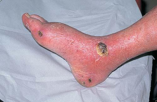
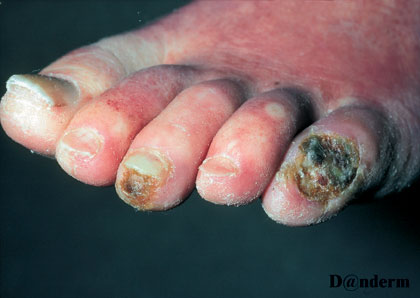
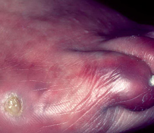
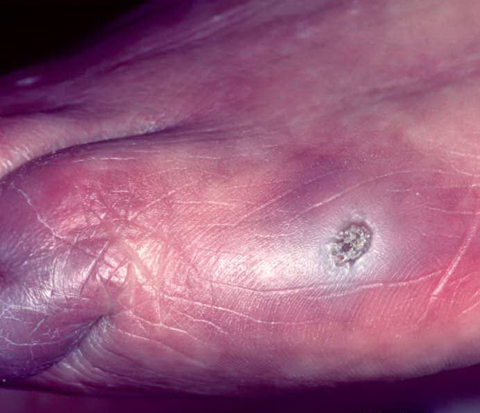
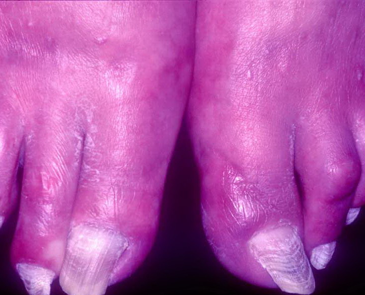
PresentationChronic leg or vascular ulcers typically manifest as arterial, neurotrophic, or venous ulcers. They are distinct with regard to their location, appearance, bleeding, and associated pain and findings Arterial ulcers Arterial ulcers are often located distally and on the dorsum of the foot or toes. Initially they have irregular edges, but they may progress to have a better-defined appearance. The ulcer base contains grayish, unhealthy-appearing granulation tissue. With manipulation, such as debriding, these ulcers bleed very little or not at all. The patient may report characteristic pain, especially at night when supine, which is relieved by dependency of the extremity. Upon examination, characteristic findings of chronic ischemia, such as hairlessness, pale skin, and absent pulses, are noted.
|