| Carcinome spinocellulaire=السرطان شائك الخلايا |
|
| atlas of dermatology - C |
| Thursday, 07 October 2010 22:38 |
|
Cutaneous squamous cell carcinoma Cutaneous squamous cell carcinoma (SCC) is the second most common form of skin cancer and accounts for 20% of cutaneous malignancies.1 Squamous cell carcinoma frequently arises on the sun-exposed skin of middle-aged and elderly individuals. Most squamous cell carcinomas are readily identified and removed in the physician's office as a minor surgical procedure. Larger and more invasive lesions may require aggressive surgical management, radiation therapy, or both. High-risk squamous cell carcinoma carries a significant risk of metastasis and, as such, requires careful evaluation and treatment. An estimated 8000 cases of nodal metastasis and 3000 deaths occur in the United States annually, almost wholly attributable to aggressive or high-risk squamous cell carcinoma.2,3
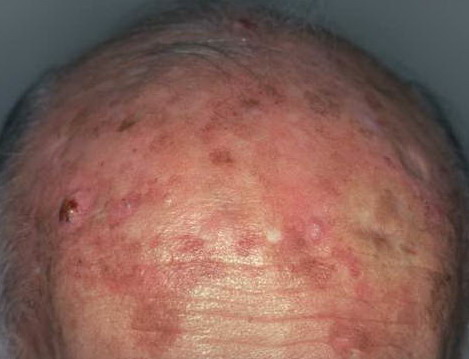
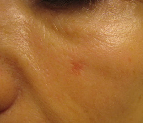
PathophysiologySquamous cell carcinoma (SCC) is a malignant tumor of epidermal keratinocytes. Some cases of squamous cell carcinoma occur de novo (ie, in the absence of a precursor lesion); however, some squamous cell carcinomas arise from sun-induced precancerous lesions known as actinic keratoses. Patients with multiple actinic keratoses are at increased risk for developing squamous cell carcinoma.12 Squamous cell carcinoma is capable of locally infiltrative growth, spread to regional lymph nodes, and distant metastasis, most often to the lungs. A detailed patient history often reveals the presence of one or more risk factors for squamous cell carcinoma (SCC) (see general risk factors in Background). Most squamous cell carcinomas are discovered by patients and are brought to a physician's attention by the patient or a relative. The typical squamous cell carcinoma manifests as a new or enlarging lesion that concerns the patient. Squamous cell carcinoma is typically a slow-growing malignancy, but some lesions enlarge rapidly. Although most squamous cell carcinoma patients are asymptomatic, symptoms such as bleeding, weeping, pain, or tenderness may be noted, especially with larger tumors. Numbness, tingling, or muscle weakness may reflect underlying perineural involvement, and this history finding is important to elicit because it adversely impacts prognosis.19 Actinically derived squamous cell carcinoma The most common type of squamous cell carcinoma is the sun-induced type. As such, a history of long-term sun exposure dating back to childhood is frequently elicited. Many patients report having experienced multiple blistering sunburns during their lifetime, while others may have used indoor tanning beds or received UV light therapy (eg, psoralen plus UVA [PUVA] for psoriasis). Patients may have been treated in the past for sun-induced lesions such as actinic keratoses, basal cell carcinoma, melanoma, or squamous cell carcinoma. Immune suppression Patients should always be questioned about possible sources of immunosuppression. A history of solid-organ transplantation, hematologic malignancy (particularly chronic lymphocytic leukemia), HIV infection or AIDS, or long-term use of immunosuppressive medications (eg, as treatment for an autoimmune condition) may be elicited. Marjolin ulcer This eponym most frequently refers to a squamous cell carcinoma that arises from chronically scarred or inflamed skin; however, malignant transformation to a basal cell carcinoma, melanoma, or sarcoma may also occur.20 Patients may report a change in the skin (eg, induration, elevation, ulceration, weeping) at the site of a preexisting scar or ulcer. The average latency period is 35 years21 ; therefore, the diagnosis requires a high index of clinical suspicion. Marjolin ulcers are associated with a high rate of metastasis, estimated at 30%,22,23 and a mortality rate of 33%.24 HPV-associated squamous cell carcinoma Virally induced squamous cell carcinoma most commonly manifests as a new or enlarging warty growth on the penis, vulva, perianal area, or periungual region. Patients often present with a history of "warts" that have been refractory to various treatment modalities in the past. A history of previously documented genital HPV infection may be elicited. PhysicalSquamous cell carcinoma (SCC) may manifest as a variety of primary morphologies, with or without associated symptoms. Note the following: Squamous cell carcinoma in situ Squamous cell carcinoma in situ is defined histologically by atypia involving the full thickness of the epidermis but without invasion into the dermis. Clinically, lesions of squamous cell carcinoma in situ range from a scaly pink patch to a thin keratotic papule or plaque similar to an actinic keratosis. Bowen disease is a subtype of squamous cell carcinoma in situ characterized by a sharply demarcated pink plaque arising on non–sun-exposed skin. Erythroplasia of Queyrat refers to Bowen disease of the glans penis, which manifests as one or more velvety red plaques.
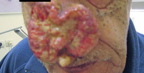
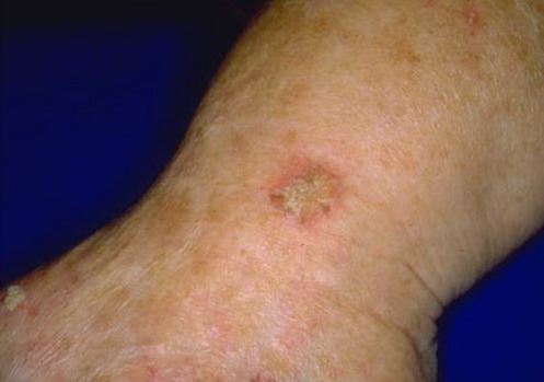
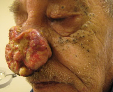
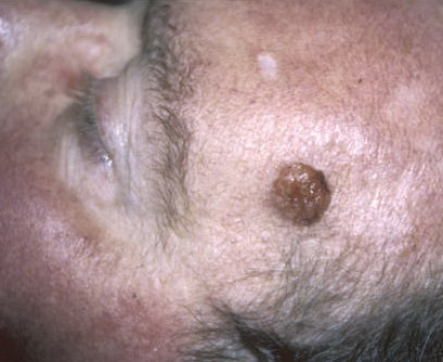
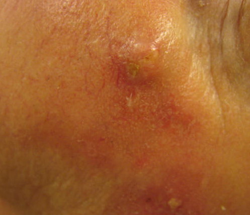
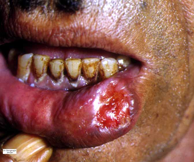
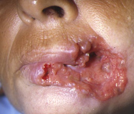
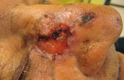
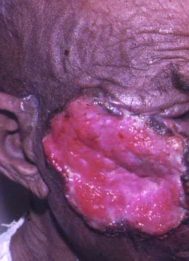
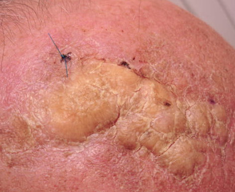
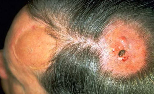
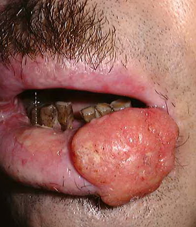
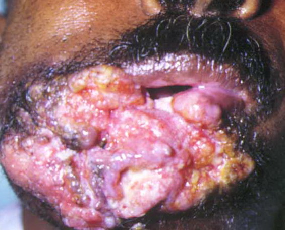
Typical squamous cell carcinoma The characteristic invasive squamous cell carcinoma is a raised, firm, pink-to-flesh–colored keratotic papule or plaque arising on sun-exposed skin. Approximately 70% of all squamous cell carcinomas occur on the head and neck, with an additional 15% found on the upper extremities. Surface changes may include scaling, ulceration, crusting, or the presence of a cutaneous horn. Less commonly, squamous cell carcinoma may manifest as a pink cutaneous nodule without overlying surface changes. The absence of surface changes should raise suspicion of a metastatic focus from another skin or nonskin primary site or a different and potentially more lethal tumor such as Merkel cell carcinoma. A background of severely sun-damaged skin, including solar elastosis, mottled dyspigmentation, telangiectasia, and multiple actinic keratoses, is often noted
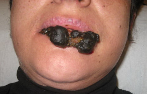
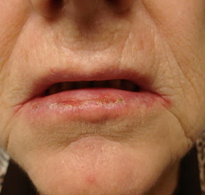
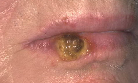
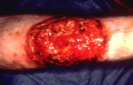
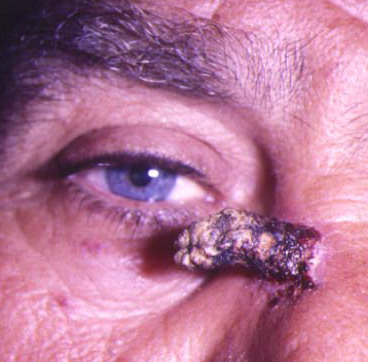
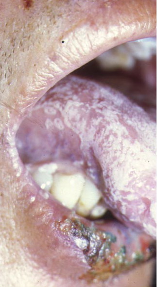
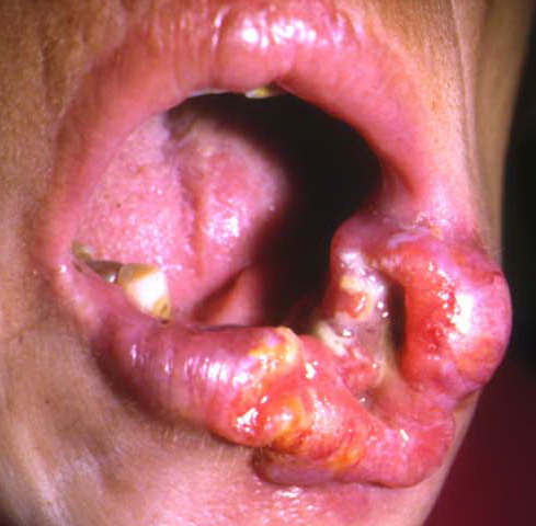
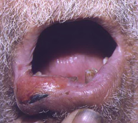
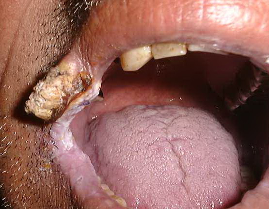
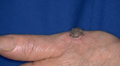
Periungual squamous cell carcinoma Periungual squamous cell carcinoma typically mimics a verruca and is frequently misdiagnosed for years as a wart prior to biopsy. Less commonly, lesions may resemble chronic paronychia with swelling, erythema, and tenderness of the nail fold; onychodystrophy also may be noted. Periungual squamous cell carcinomas are frequently associated with HPV.25 Marjolin ulcer This subtype of squamous cell carcinoma appears as a new area of induration, elevation, or ulceration at the site of a preexisting scar or ulcer. Patients with this form of squamous cell carcinoma can have a poor prognosis. The diagnosis of Marjolin ulcer should be considered in any ulcer that fails to heal with standard therapy. Perioral squamous cell carcinoma Squamous cell carcinoma of the lip usually arises on the vermillion border of the lower lip. It is sometimes predated by a precursor lesion, actinic cheilitis, which manifests as xerosis, fissuring, atrophy, and dyspigmentation. Actinic cheilitis is analogous to actinic keratosis of the skin. Squamous cell carcinoma on the lip manifests as a new papule, erosion, or focus of erythema/induration. Intraoral squamous cell carcinoma typically manifests as a white plaque (leukoplakia) with or without reddish reticulation (erythroplakia). Common locations include the anterior floor of the mouth, the lateral tongue, and the buccal vestibule. Anogenital squamous cell carcinoma Squamous cell carcinoma in the anogenital region may manifest as a moist, red plaque on the glans penis; indurated or ulcerated lesions may be seen on the vulva, external anus, or scrotum. Associated symptoms include pain, pruritus, and intermittent bleeding. These squamous cell carcinomas are also associated with HPV infection. Verrucous carcinoma Verrucous carcinoma is a subtype of squamous cell carcinoma that can be locally destructive but rarely metastasizes. Lesions appear as exophytic, fungating, verrucous nodules or plaques, which may be described as cauliflowerlike. Verrucous carcinoma is further subdivided based on its location in the anogenital region (Buschke-Löwenstein tumor), the oral cavity (oral florid papillomatosis), and the plantar foot (epithelioma cuniculatum). Lymphadenopathy With any invasive (not in situ) squamous cell carcinoma, regional lymph nodes should be examined. Lymph node enlargement must be further evaluated by fine-needle aspiration (FNA) or nodal biopsy. CausesThe primary cause of most squamous cell carcinoma (SCC) is cumulative lifetime sun exposure. The frequency of squamous cell carcinoma is increased at lower latitudes, correlating with an increased intensity of ambient light. Other causes of squamous cell carcinoma are discussed below. UV sunlight exposure The component of sunlight believed to be most important in cutaneous carcinogenesis is UVB (290-320 nm), which is both an initiator and a promoter of carcinogenesis. In animal models, UV-induced photocarcinogenesis appears to involve the UVB and UVA-2 spectral ranges.26 UVB-induced photocarcinogenesis appears to work by suppressing the immune system in several ways. The UVB spectrum inhibits antigen presentation, induces the release of immunosuppressive cytokines, and elicits DNA damage, specifically the generation of pyrimidine dimers in keratinocyte DNA that is a molecular trigger of UV-mediated immunosuppression.27 Inactivation of the tumor suppressor gene TP53 occurs in up to 90% of all cutaneous squamous cell carcinoma lesions.28 Other tumor suppressor genes found to be mutated in squamous cell carcinoma include P16 (INK4a) and P14 (ARF).29 Therapeutic UV exposure UV light treatments used for psoriasis (and other recalcitrant dermatoses) also predispose to the development of squamous cell carcinoma. PUVA is particularly phototoxic and mutations in both TP53 and the oncogene Ha-Ras are present in a large proportion of PUVA-associated squamous cell carcinoma.30 In addition to being mutagenic, UVA in conjunction with UVB is a potent suppressor of the cutaneous immune system, which likely contributes to its role in cutaneous carcinogenesis. Fair complexion Individuals with skin types I and II account for most of the patients who develop squamous cell carcinoma; patients with oculocutaneous albinism are also at risk, and squamous cell carcinomas account for the most common type of cutaneous malignancy in this group. Such individuals lack natural protection from UV-induced carcinogenesis, owing to reduced levels of the photoprotective pigment, melanin.31 Ionizing radiation Therapeutic ionizing radiation is typically associated with the later development of basal cell carcinomas, but the risk of developing squamous cell carcinomas is also increased.32 Most patients with radiation-induced tumors have a remote history of x-ray therapy for acne vulgaris, although patients developing squamous cell carcinoma in radiation ports for Hodgkin disease or thyroid cancer treatment is not uncommon. Chemical carcinogens Exposure to arsenic is a well-established cause of cutaneous squamous cell carcinoma and internal cancers.6 Today, the main source of arsenic is contaminated well water, although arsenic may also be found in traditional Chinese medicines. Other carcinogens associated with squamous cell carcinoma include polycyclic aromatic hydrocarbons such as tar, soot, and pitch. DNA repair failure Healthy human skin is constantly repairing UV-induced damage through DNA repair mechanisms. Patients with xeroderma pigmentosum have a deficiency in an enzyme essential for normal DNA repair and are thus prone to the development of innumerable squamous cell carcinomas, and, less commonly, other cutaneous tumors.33 Iatrogenic immunosuppression The use of immunosuppressive medications to prevent rejection in organ transplant recipients is associated with a 65- to 250-fold increased risk of developing squamous cell carcinoma compared with the general population.34 The primary risk factor in these patients is cumulative lifetime UV exposure in combination with having Fitzpatrick skin type I or II. This risk also increases with the number of years post-transplantation, presumably because of the cumulative effects of prolonged immunosuppressive therapy. The greatest risk occurs in heart transplant patients, with diminishing risk seen in recipients of kidney and liver transplants, which correlates with the degree of immunosuppression (ie, number and/or dosage of medications) typically required to prevent rejection in these patient populations. Pretransplantation end-organ disease may also impact the development of post-transplant squamous cell carcinoma. For example, among renal transplant recipients, the highest prevalence of skin cancer was observed in patients with polycystic kidney disease, while the lowest incidence was seen in those with diabetic nephropathy. Similarly, cholestatic liver disease was associated with a greater post-transplantation risk of skin cancer compared with other causes of liver failure. Noniatrogenic immunosuppression In addition to iatrogenic immunosuppression, defects in cell-mediated immunity related to lymphoproliferative disorders (eg, chronic lymphocytic leukemia) predispose to the development of aggressive squamous cell carcinoma. The specific mechanisms by which immunosuppression leads to squamous cell carcinoma development are poorly understood, but diminished immunosurveillance is thought to be critical. CD8+ T cells specific for the tumor suppressor gene TP53 have been observed in patients with squamous cell carcinoma, suggesting that a functional immune system may target keratinocytes expressing mutated TP53.35 Suppression of the immune system would presumably abrogate this response and might be expected to facilitate the development of squamous cell carcinoma. Human papillomavirus Infection with specific subtypes of HPV is believed to play a role in the development of anogenital and periungual squamous cell carcinoma. Attempts to definitively link squamous cell carcinoma to HPV have yielded contradictory results, as most squamous cell carcinomas tumors outside of anogenital and periungual sites do not contain HPV. The International Agency for Research on Cancer (IARC; Lyon, France) has determined that current evidence only supports HPV types 5 and 8 as possible carcinogens.36 However, HPV types 6 and 11 have been associated with Buschke-Löwenstein tumors, whereas HPV type 16 has been frequently identified in both genital and periungual squamous cell carcinoma, suggesting the possibility of genital-digital spread.37,38 HPV types 5 and 8 have been associated with cutaneous squamous cell carcinoma in the setting of epidermodysplasia verruciformis and some solid organ transplant patients.39 Chronic inflammation Chronic inflammation, irrespective of the underlying etiology, may lead to the development of squamous cell carcinoma. Both noninfectious inflammatory diseases and chronic infections have been associated with squamous cell carcinoma. Likewise, the Marjolin ulcer variant of squamous cell carcinoma may develop in patients with a chronic scarring condition such as dystrophic epidermolysis bullosa. In fact, the leading cause of death in patients with dystrophic epidermolysis bullosa is metastatic cutaneous squamous cell carcinoma,10 with an 80% mortality rate within 5 years of diagnosis of squamous cell carcinoma40 and with two thirds of patients dying from metastatic disease.41 More recently, evidence suggests that patients with junctional epidermolysis bullosa may also be at increased risk for developing squamous cell carcinoma.42 The underlying pathogenesis of such lesions is not understood, but mutations in the TP53 and P16 tumor suppressor genes have been described in dystrophic epidermolysis bullosa–associated squamous cell carcinoma.43 Conditions that predispose to the development of squamous cell carcinoma Chronic inflammatory and scarring conditions are as follows:
Chronic infections are as follows:
Genetic syndromes and dermatoses are as follows:
Imaging is not routinely indicated for diagnosing cutaneous (SCC). However, radiologic imaging should be obtained in patients with regional lymphadenopathy and/or neurologic symptoms suggestive of perineural involvement, for nodal staging, and for preoperative planning in patients in whom deep or extensive tissue involvement is suspected. CT scanning, MRI, ultrasonography, or positron-emission tomography (PET) scanning may be used depending on the specific question being addressed, although the selection of one modality over another is often based on clinician and institutional preference. Currently, no formal guidelines have been developed regarding the use of radiologic imaging in cutaneous squamous cell carcinoma. Physical examination of lymph nodes In all squamous cell carcinoma patients, the draining nodal basins should be palpated. If nodes are palpable, a biopsy should be performed using fine-needle aspiration (FNA) or excision. If lymph nodes are clinically negative but the tumor meets high-risk criteria, little data are available to guide what should be done next. Subsequently, management currently varies with regard to further staging.44 See "High-risk squamous cell carcinoma" in Prognosis. Radiologic staging Only a few studies have reported on the utility of radiologic imaging in cutaneous squamous cell carcinoma. One study of MRI and CT scanning in patients with histologically proven perineurally invasive squamous cell carcinoma showed that only 20% of asymptomatic patients have positive findings discovered from imaging studies. Thus, CT scanning and MRI appear to be poor in detecting asymptomatic nerve involvement. However, positive imaging findings did correlate with worse outcomes. The 5-year survival rate was 50% if CT scanning or MRI findings were positive, versus 86% if they were negative.45 Two studies reported on radiologic imaging for detecting subclinical nodal metastasis.46 The first, a study of vulvar squamous cell carcinoma, indicated that ultrasonography followed by FNA for suspicious nodes was superior to CT scanning in staging subclinical nodal metastasis, with ultrasound-guided FNA demonstrating 80% sensitivity and 100% specificity. The second is a small study of PET scanning in 9 patients with high-risk squamous cell carcinoma. PET scanning detected subclinical nodal metastasis in 3 of 9 patients.47 Thus, PET scanning and ultrasound-guided FNA may be capable of detecting many cases of subclinical nodal metastasis. Sentinel lymph node biopsy Summary Little data are available to guide decisions about staging of nodal basins in high-risk squamous cell carcinoma. However, PET scanning, ultrasound-guided FNA, and SLNB all appear to offer a good chance of detecting subclinical nodal metastasis with low morbidity. Thus, nodal staging may be considered in patients with high-risk squamous cell carcinoma. Development of prognostic models that better predict the risk of nodal metastasis will allow for more rational decisions about which patients should undergo nodal staging. ProceduresSkin biopsy Histologic FindingsThe biopsy report for squamous cell carcinoma (SCC) often carries prognostic implications. Recognizing the implications of the various histologic subtypes of squamous cell carcinoma is important, and the astute clinician uses his or her understanding of histopathology to advantage in planning the appropriate therapeutic intervention.
StagingSquamous cell carcinoma (SCC) is staged according to American Joint Committee on Cancer (AJCC) guidelines, which use the tumor, node, metastasis (TNM) classification system.49 This staging system has been updated to incorporate information about tumor factors that impact prognosis. Cutaneous squamous cell carcinoma of the eyelid is excluded from the updated system.
The new TNM staging system has also revised nodal staging. Previously, the system had only had a single N1 level to signify nodal involvement. The new system has 5 levels. The decision to stage patients according to extent of nodal disease was based on significant findings of several studies, both prospective and retrospective, showing that the number and size of lymph node involvement correlated with patient prognosis.50,51,52,53,54 In the new staging system, N1 disease involves a single ipsilateral node 3 cm or smaller in its largest dimension. N2a disease includes cases with a single ipsilateral node greater than 3 cm but less than or equal to 6 cm. N2b refers to those with multiple ipsilateral nodes smaller than or equal to 6 cm. N2c includes cases of bilateral or contralateral involvement less than or equal to 6 cm. N3 disease is reserved for cases with any involved node greater than 6 cm. In early 2010, Milross et al proposed an alternative nodal staging system (the N1S3), which also stages cutaneous squamous cell carcinoma patients based on the number (single or multiple) and size (< or > 3 cm) of lymph nodes involved, but also incorporates the parotid as one of the regional levels. Stage I disease refers to those with a single lymph node measuring less than or equal to 3 cm. Stage II includes cases with a single lymph node greater than 3 cm or multiple lymph nodes less than or equal to 3 cm, while stage III is any patient with multiple lymph nodes greater than 3 cm.55 This system was found to have a significant predictive capacity for locoregional control (P <.0001), disease-specific survival (P <.0001), and overall survival (P <.0001) in a group of 215 patients and was reproduced on external validation in a cohort of 250 patients. Distant metastases are staged according to the presence (M1) or absence (M0) of metastases in distant organs or sites outside of regional lymph nodes. This remains unchanged from the previous TNM staging system Stage 0 is equivalent with in situ disease. Disease stages I and II include patients with T1 and T2 tumors, respectively who have no nodal or distant metastasis (N0, M0). Stage III disease includes T3 cases without nodal involvement (N0) or cases with N1 involvement. Stage IV includes those with T4 disease, or N2 or N3 disease, or distant metastasis (M1).
Medical CareNonsurgical options for the treatment of cutaneous squamous cell carcinoma (SCC) include topical chemotherapy, topical immune response modifiers, photodynamic therapy (PDT), radiotherapy, and systemic chemotherapy. The use of topical therapy and PDT is generally limited to premalignant (ie, actinic keratoses) and in situ lesions. Radiation therapy is a primary treatment option for patients in whom surgery is not feasible and is an adjuvant therapy for those with metastatic or high-risk cutaneous squamous cell carcinoma. In current practice, systemic chemotherapy is used exclusively for patients with metastatic disease. However, newer, more targeted drugs, such as epidermal growth factor receptor (EGFR) antagonists (eg, cetuximab), have favorable adverse effect profiles and await trails to determine if they are beneficial in high-risk squamous cell carcinoma. Surgical CareMost squamous cell carcinomas (SCCs) are readily treated in the physician's office by surgical or destructive methods, with a high expectation of cure. The treatment of squamous cell carcinoma must take into account multiple patient- and lesion-specific factors. The standard modalities available for the treatment of localized (primary) invasive squamous cell carcinoma are described below. Because squamous cell carcinoma is a lesion that can recur, metastasize, and cause death, and, because recurrent squamous cell carcinoma carries a worse prognosis, every opportunity should be taken to effect complete tumor extirpation at first presentation.
|
| Last Updated on Saturday, 05 February 2011 07:04 |