| Venous lake = بحيرة وريدية |
|
| atlas of dermatology - V |
| Wednesday, 27 October 2010 14:37 |
|
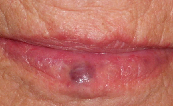
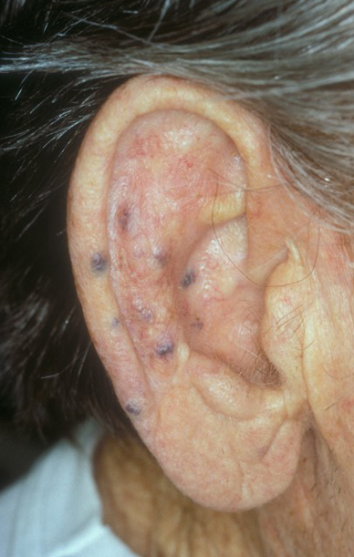
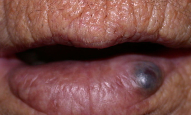
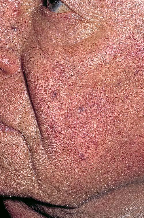
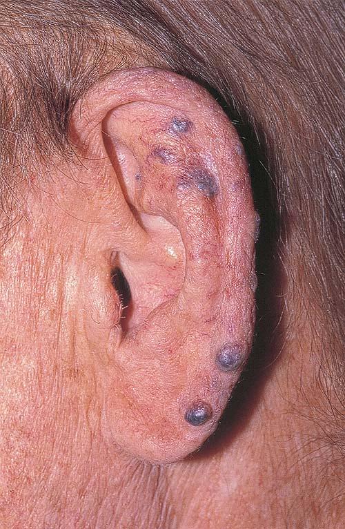
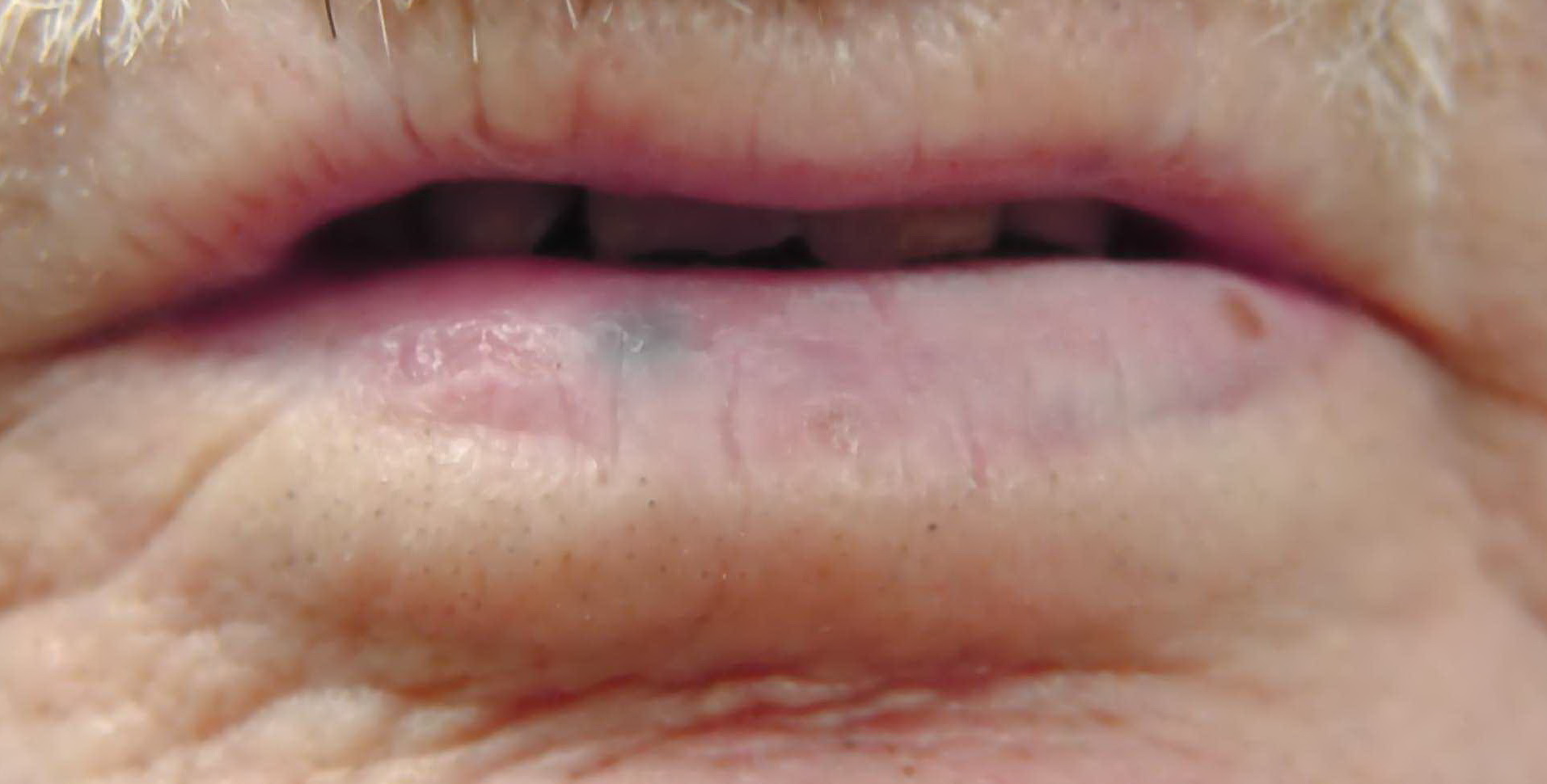
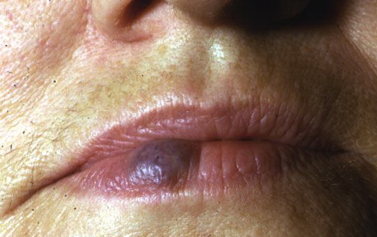
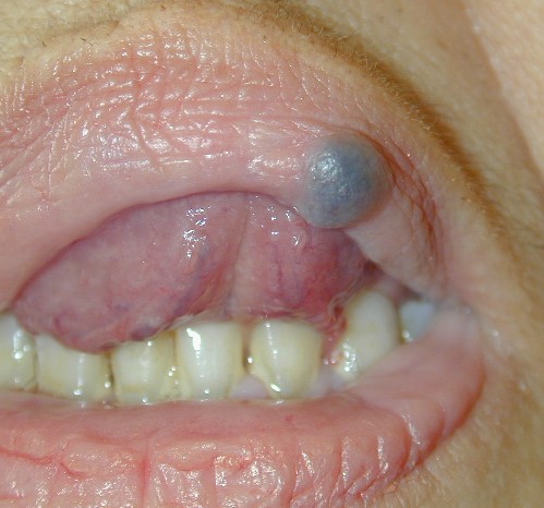
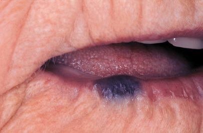
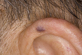
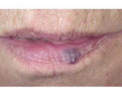
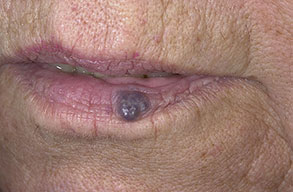
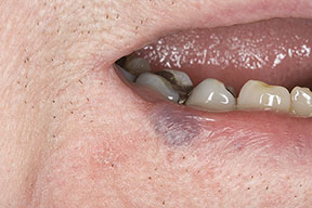
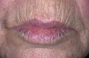
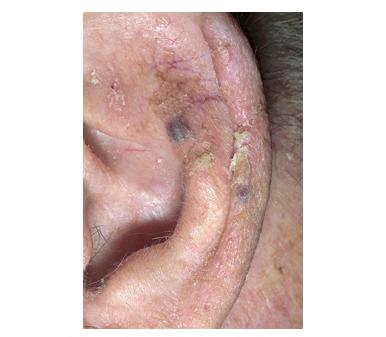
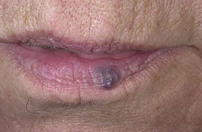
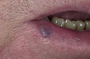
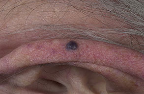
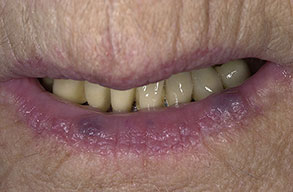
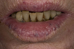
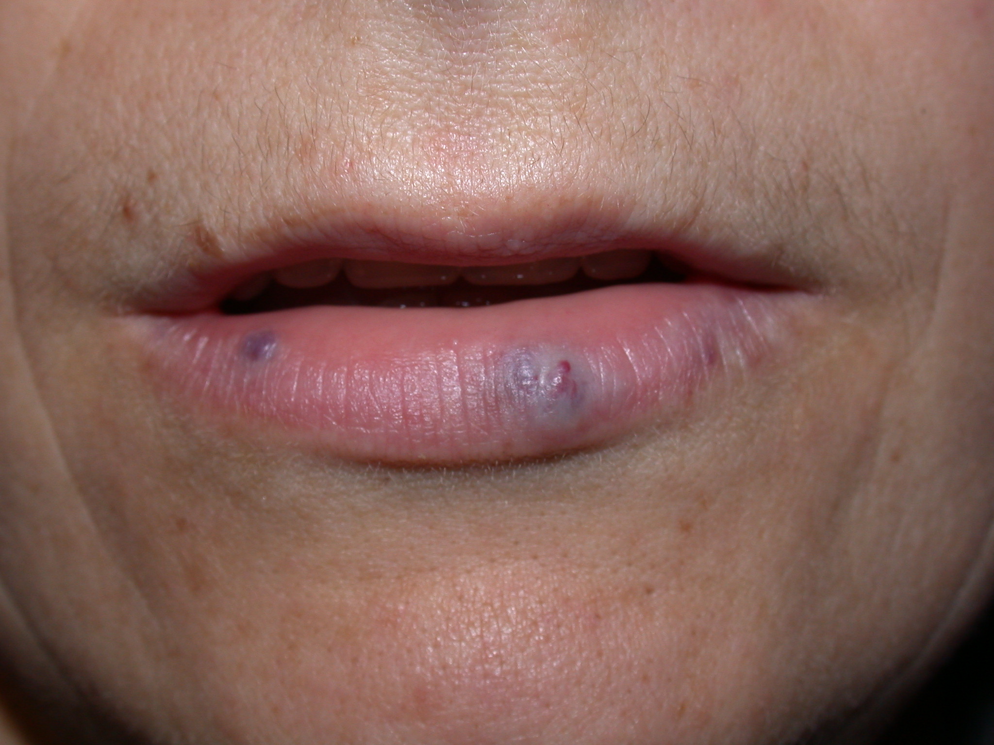
Venous Lakes Venous lakes manifest as dark blue-to-violaceous compressible papules caused by dilation of venules. They were first described in 1956 by Bean and Walsh, who noted their compressibility and predilection for sun-exposed skin, especially the ears of elderly patients.1 Although benign, venous lakes are important because of their mimicry of malignant lesions, such as melanoma and pigmented basal cell carcinoma. A venous lake is an acquired form of vascular ectasia (vascular dilatation). A capillary aneurysm is considered a precursor or variant of a venous lake. The exact incidence of venous lakes is unknown but they are believed to be common. The worldwide incidence of venous lakes is unknown but is believed to be the same as that in the Mortality from venous lakes has not been reported. Venous lakes are usually asymptomatic, although pain, tenderness, and excessive bleeding may occur if a lesion is traumatized. Venous lakes are considered biologically harmless. No racial predilection has been documented for venous lakes. Bean and Walsh reported that 95% of venous lakes were observed in males.1 Another review of venous lakes confirmed the same sex distribution. The disproportionate male distribution may be related to occupational sun exposure, hair length, and hairstyles. Women comprised the majority of treated patients in a large study of laser therapy for venous lakes; however, this may be related to increased concern among women regarding cosmetic appearance rather than with true incidence. Venous lakes have been reported only in adults and usually occur in patients older than 50 years. The average age of presentation for venous lakes has been reported to be 65 years. Venous lakes most commonly occur in adults older than 50 years with a history of long-term sun exposure. The typical presentation is a slow-growing asymptomatic lesion. Patients with venous lakes may report that the papule has been present for several years prior to presentation. Recurrent bleeding after minor trauma may also be reported. Physical examination usually reveals a soft, compressible, dark-blue or violaceous papule (slightly elevated lesion), up to 1 cm in diameter. Venous lakes usually are well demarcated, with a smooth surface. Compression often causes a emptying of the blood content. Venous lakes typically are distributed on the sun-exposed surfaces of the face and neck, especially on the helix and antihelix of the ear and the posterior aspect of the pinna, as shown in the image below. Another common site of involvement is the vermilion border of the lower lip, shown below. Sometimes, several lesions are found on the same person, and the surrounding skin reveals actinic damage, as shown below. Two main theories regarding the development of venous lakes have been proposed. The first involves injury to the vascular adventitia and the dermal elastic tissue due to long-term solar damage permitting dilatation of superficial venous structures. The second theory involves the involvement of vascular thrombosis in the development of venous lakes. Thrombosis is commonly present in lesions of this type; however, whether the thromboses is a primary or a secondary event in the development of these lesions is unclear.
Treatment
Surgical Care
Drugs cannot be used to ameliorate or remove venous lakes. |
| Last Updated on Saturday, 04 December 2010 11:50 |